Cervical caner image automatic partition method based on T2-magnetic resonance imaging (MRI) and dispersion weighted (DW)-MRI

A T2-MRI, T2-MR technology, applied in the field of image processing, can solve the problems of intensity overlap, regional growth segmentation of tumors, blurred tumor boundaries, etc., to achieve the effect of overcoming noise

Active Publication Date: 2013-03-27
INST OF AUTOMATION CHINESE ACAD OF SCI
View PDF2 Cites 11 Cited by
  • Summary
  • Abstract
  • Description
  • Claims
  • Application Information

AI Technical Summary

Problems solved by technology

from figure 1 (a) It can be seen that the T2-MR image has a relatively high spatial resolution and the tumor border is relatively clear, but the normal tissue of the cervix, tumor, bladder wall and rectum have serious intensity overlap; from figure 1 (b) It can be seen that the tumor has a significantly

Method used


Image

  • Cervical caner image automatic partition method based on T2-magnetic resonance imaging (MRI) and dispersion weighted (DW)-MRI
  • Cervical caner image automatic partition method based on T2-magnetic resonance imaging (MRI) and dispersion weighted (DW)-MRI
  • Cervical caner image automatic partition method based on T2-magnetic resonance imaging (MRI) and dispersion weighted (DW)-MRI

Examples


Embodiment Construction

[0014] In order to make the object, technical solution and advantages of the present invention clearer, the present invention will be described in further detail below in conjunction with specific embodiments and with reference to the accompanying drawings.
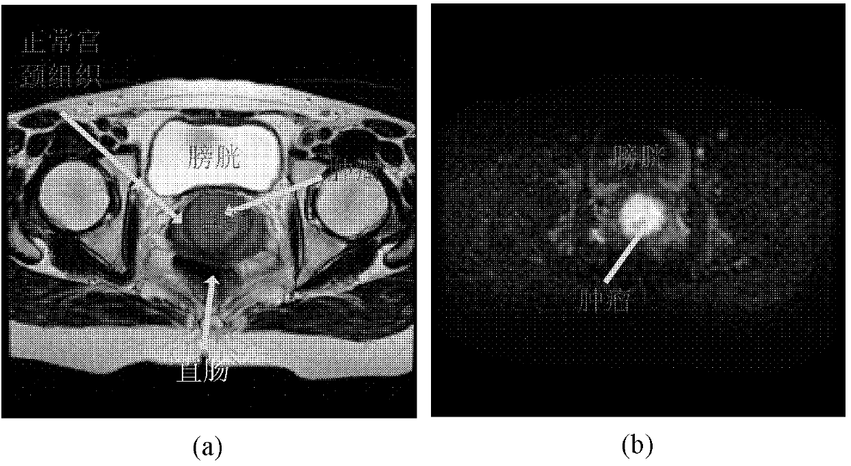
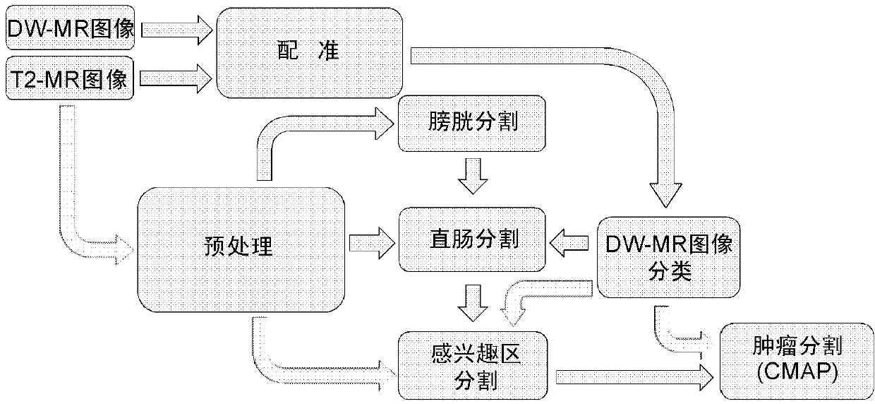
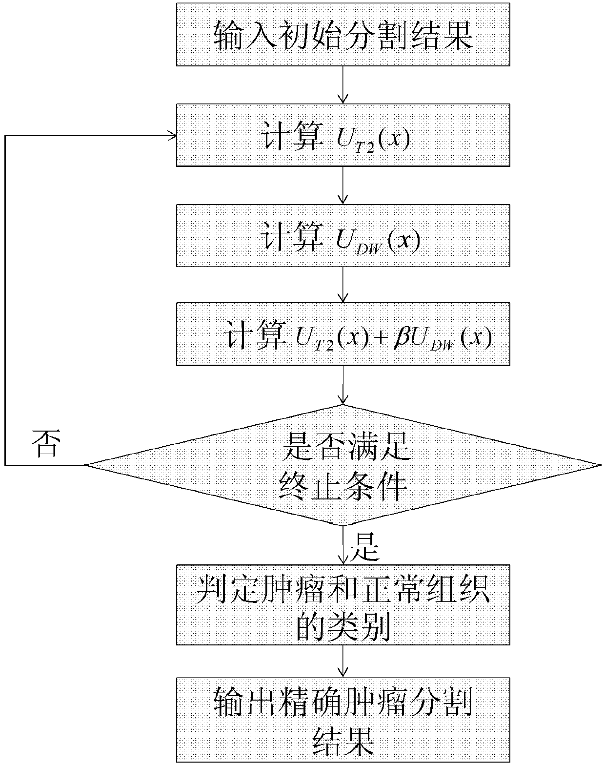
[0015] The core idea of ​​the present invention is an automatic segmentation framework for cervical cancer images based on T2-weighted MRI (T2-MRI) and diffusion-weighted MRI (DW-MRI) and the use of joint maximum a posteriori probability (CMAP) to accurately The method for segmenting the tumor region of cervical cancer, the specific steps include: first, use the non-linear registration method to register the DW-MR image to the T2-MR image (the mutual information registration method is used as an example here), and the registered The DW-MR images are classified; then the T2-MR images are filtered using the nonlinear anisotropic diffusion filtering technique (here the P-M nonlinear anisotropic diffusion filtering is used as ...


PUM


Abstract

A cervical caner image automatic partition method based on T2 weighted magnetic resonance imaging (MRI) T2-MRI and dispersion weighted (DW)-MRI includes that a DW-MR image is registered to a T2-MR image by using a non-linear register method, and the registered DW-MR image is sorted; the T2-MR image is filtered through non-linear anisotropic diffusion filtering technology, a bladder and a rectum are partitioned, and an interested area is partitioned through a partition result of the bladder and the rectum; and a combined maximum a posterior (CMAP) method is adopted for an interested area of the T2-MR image and the DW-MR image to conduct precise partition of a tumor. The cervical caner image automatic partition method fully uses effective information of the T2-MR image and the DW-MR image, can effectively overcome effects of noise, partial volume effect and strength overlapping in the T2-MR image, is precise and effective, and has important clinical and application value on prevention, diagnosis and treatment of the cervical cancer.

Description

technical field [0001] The invention belongs to the field of image processing, and in particular relates to an automatic segmentation method for cervical cancer images based on T2-weighted nuclear magnetic resonance imaging T2-MRI and diffusion-weighted nuclear magnetic resonance imaging DW-MRI. Background technique [0002] Cervical cancer is one of the common malignant tumors that seriously threaten women's life and health. Accurate segmentation of cervical cancer has important clinical significance and application value for the prevention, diagnosis and treatment of cervical cancer. [0003] With the development of imaging technology, medical image segmentation has become a key and challenging problem in the field of medical image analysis. Cervical cancer segmentation is particularly complex due to the complex structure of human abdominal tissue, and a single imaging mode T2-MRI cannot fully display the effective information of cervical cancer. Such as figure 1 (a) an...

Claims


Application Information

Patent Timeline
IPC IPC(8): G06T7/00
CPCG06T7/00G06T7/143G06T7/11G06T2207/10088G06T2207/20076G06T2207/30004
Inventor 李悟考月英田捷
Owner INST OF AUTOMATION CHINESE ACAD OF SCI
Why Eureka
Try Eureka