Eureka AIR delivers breakthrough ideas for toughest innovation challenges, trusted by R&D personnel around the world.

a percutaneous nephroscope

A technology of nephroscope and mirror bridge, which is applied in the field of percutaneous nephroscope, can solve the problems of easily torn calices, increase patient injury, and make adjustment difficult, so as to reduce the risk of tearing kidney tissue, reduce tissue damage, and save energy. The effect of operation time

Active Publication Date: 2016-05-11
中山市环能缪特斯医疗器械科技有限公司
View PDF8 Cites 4 Cited by
  • Summary
  • Abstract
  • Description
  • Claims
  • Application Information

AI Technical Summary

Problems solved by technology

[0009] 1. When performing puncture under the guidance of B-ultrasound, due to the artifacts and partial volume effects of ultrasound, doctors are often unable to determine whether the needle tip has reached the target position;
[0010] 2. When performing puncture under the guidance of X-ray (C-arm machine), it is only possible to judge whether the needle tip has reached the target position through the plane image of X-ray, which depends on the operator's surgical experience, and both doctors and patients are subject to radiation; the number of actions Multiple, puncture needle and puncture sheath insertion, withdrawal of puncture needle, insertion of guide wire, withdrawal of puncture sheath, insertion of dilator, dilation operation, withdrawal of dilator, insertion of percutaneous nephroscope, extraction of guide wire, insertion of surgical instruments, positioning 1. Performing 12 actions such as surgery, the operation time is long, the patient is greatly injured, and the operator's work intensity is increased, resulting in fatigue;
[0011] 3. Every operation must expand the puncture hole, and the expansion will increase the harm to the patient;
[0012] 4. The existing percutaneous nephroscope has a large diameter (outer diameter is 12 or 18 Fr, equivalent to 4-6 mm), and it is difficult to adjust the position, which is easy to tear the calices, resulting in increased bleeding and even surgical failure; although it is already A minimally invasive surgery, but the wound is still too large and difficult to heal

Method used


Image

  • a percutaneous nephroscope
  • a percutaneous nephroscope
  • a percutaneous nephroscope

Examples


Embodiment 1

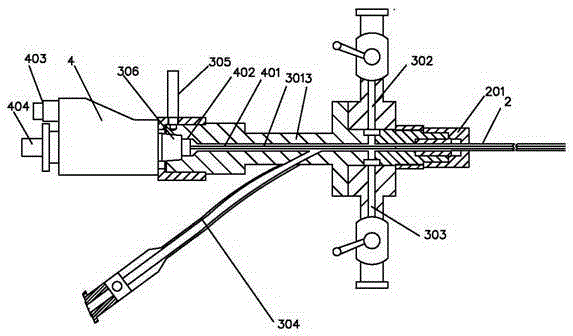
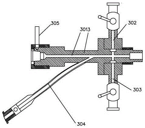
[0046] A percutaneous nephroscope, such as Figure 1 to Figure 4 As shown, it includes a mirror bridge assembly, a puncture needle 1, a mirror sheath assembly and a lens 401 assembly, the mirror bridge assembly is provided with a mirror bridge channel 301 and a mirror bridge seat 3, and the mirror sheath assembly is provided with a mirror sheath channel 2 and The mirror sheath seat 201 arranged at the end of the mirror sheath channel 2,

[0047] When performing the puncture step, the front end of the puncture needle 1 is inserted into and protrudes from the sheath channel 2, and the end of the puncture needle 1 abuts against the sheath seat 201;

[0048] When the mirror sheath base 201 is sealed and connected with the mirror bridge base 3, the mirror sheath channel 2 is connected with the mirror bridge channel 301, and the lens 401 assembly passes through the mirror bridge channel 301 and the mirror bridge channel 301 in turn. Mirror sheath channel 2.

[0049] 1. Simplify th...


PUM


Abstract

A percutaneous nephroscope comprises a bridge assembly, a puncture needle, a sheath assembly and a lens assembly, wherein the bridge assembly is provided with a bridge passage and a bridge base, and the sheath assembly is provided with a sheath passage and a sheath base arranged on the end of the sheath passage. In the step of puncture, the front end of the puncture needle is inserted into and extends out of the sheath passage, and the end of the puncture needle abuts against the sheath base; when the sheath base is tightly connected with the bridge base, the sheath passage communicates with the bridge passage, and the lens assembly sequentially passes through the bridge passage and the sheath passage. The percutaneous nephroscope has the following advantages: (1) the operating procedure is simplified, the time of an operation can be shortened, the requirement on the endurance of patients is decreased, and the working intensity of doctors can be decreased; (2) particularly, unnecessary expansion which can cause the pain and tissue injuries of patients is reduced; (3) because the diameter of the sheath is small, the percutaneous nephroscope can be flexibly operated in the narrow spaces of the renal pelvis and the renal calyx, the risk of tearing kidney tissues is reduced, and moreover, the surgical cut is small and can easily heal up after the operation.

Description

technical field [0001] The invention relates to the field of medical instruments, in particular to a percutaneous nephroscope. Background technique [0002] Minimally invasive intervention plays an increasingly important role in the examination and treatment of renal diseases such as stones and renal tumors. The current traditional percutaneous renal puncture and dilation methods are as follows: [0003] The first step is to perform the puncture, including the following steps: [0004] 1. Percutaneous renal puncture should be performed under the guidance of B-ultrasound or X-ray (C-arm machine) (now, because doctors and patients are more concerned about radiation, so the current hospital mostly uses B-ultrasound-guided puncture); 2. The puncture needle enters For renal calices, use B-ultrasound or X-ray (C-arm machine) images to confirm that the needle tip of the puncture needle has reached the target; 3. Pull out the needle core, and urine can be seen flowing out after suc...

Claims


Application Information

Patent Timeline
Patent Type & Authority Patents(China)
IPC IPC(8): A61B17/34
Inventor 谭国安
Owner 中山市环能缪特斯医疗器械科技有限公司
Why Eureka
Eureka Blog
Learn More