Electrical impedance imaging method of ultrasonic-synergy biological tissue

A biological tissue, impedance imaging technology, applied in ultrasonic/sonic/infrasound image/data processing, ultrasonic/sonic/infrasonic Permian technology, medical science, etc. problems such as poor performance, to achieve the effect of easy reconstruction and high resolution

Inactive Publication Date: 2013-06-19
CHINA JILIANG UNIV
View PDF1 Cites 11 Cited by
  • Summary
  • Abstract
  • Description
  • Claims
  • Application Information

AI Technical Summary

Problems solved by technology

[0007] 2. Less information
The so-called inherent morbidity means that the boundary voltage is not sensitive to the change of the electrical parameters in the center of the analysis area, and the morbidity of the reconstruction process means that the reconstructed image is easily affected by the error of

Method used


Image

  • Electrical impedance imaging method of ultrasonic-synergy biological tissue
  • Electrical impedance imaging method of ultrasonic-synergy biological tissue
  • Electrical impedance imaging method of ultrasonic-synergy biological tissue

Examples


Embodiment Construction

[0033] The present invention will be further described below in conjunction with the accompanying drawings and specific embodiments.

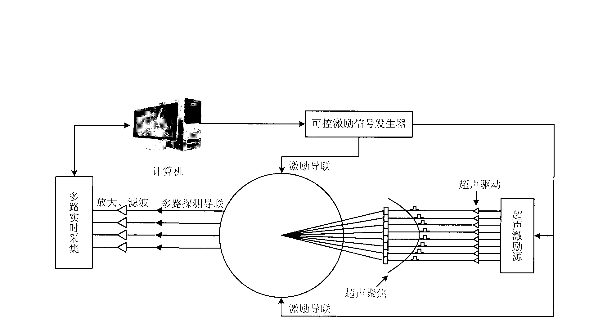
[0034] Such as figure 1 As shown, the ultrasound-assisted biological tissue electrical impedance imaging method of the present invention comprises the following steps:

[0035] (1) Several electrical excitation and detection leads are set on the surface of the biological tissue, and the controllable excitation signal generator generates the excitation signal and transmits it to the excitation lead, thereby generating electrical excitation to the biological tissue; using an ultrasonic excitation source to generate ultrasonic waves and Driven by ultrasound, the ultrasound is focused inside the biological tissue;

[0036] (2) Continuous positioning of ultrasonic waves focused on the inside of biological tissue, so as to complete the overall or partial scanning of biological tissue;

[0037] (3) Signal acquisition: Ultrasound is focused and posit...


PUM


Abstract

The invention discloses an electrical impedance imaging method of an ultrasonic-synergy biological tissue. The electrical impedance imaging method comprises that a plurality of electric excitation leads and detection guide leads are arranged on the surface of the biological tissue, and an ultrasonic excitation source is adopted to generate ultrasonic waves and the ultrasonic waves are focused in the biological tissue through ultrasonic drive; the ultrasonic waves focused in the biological tissue are located in a continuous mode, and integral or partial scanning to the biological tissue is finished; signals are collected, the ultrasonic waves are focused at a certain spatial position, and a high-frequency signal corresponding to electrical impedance of the spatial position is generated, the signals are collected through the multi-channel detection leads, amplified, filtered, and then output; after focusing and scanning of the ultrasonic waves are finished, a voltage signal after being collected is guided in a computer, and a signal is obtained according to the collected integral scanning and partial scanning to the biological tissue, and electrical impedance distribution of the biological tissue is calculated and rebuilt. Through the electrical impedance imaging method of the ultrasonic-synergy biological tissue, a high-precision and high-resolution electrical impedance image can be obtained.

Description

technical field [0001] The invention relates to the field of biological tissue imaging, in particular to an ultrasound-assisted biological tissue electrical impedance imaging method. Background technique [0002] Medical images are intuitive, vivid and informative, and play an important role in clinical diagnosis. With the advancement of medical imaging technology, medical imaging has developed from anatomical structure imaging to functional imaging, that is, medical images can not only display the morphological changes of the anatomical structure of the disease, but also reflect the functions of the body's organs and related physiological and biochemical changes, and can detect The abnormality of its physiological activities provides important information for disease diagnosis and promotes the progress and development of medicine. [0003] Traditional Electrical Impedance Tomography (Electrical Impedance Tomography, EIT) is based on the physical phenomenon that different h...

Claims


Application Information

Patent Timeline
IPC IPC(8): A61B5/053A61B8/08
Inventor 徐文龙张宁徐冰俏
Owner CHINA JILIANG UNIV
Why Eureka
Try Eureka