Current Density Imaging Method of Biological Tissue Based on Acoustoelectric Effect

A biological tissue and current density technology, applied in the direction of ultrasonic/sonic/infrasonic image/data processing, application, ultrasonic/sonic/infrasonic Permian technology, etc., can solve uncertainties, incompleteness of measurement data, inverse The problem does not have a unique solution, etc., to achieve high resolution and facilitate medical diagnosis

Inactive Publication Date: 2016-04-13
CHINA JILIANG UNIV
View PDF1 Cites 0 Cited by
  • Summary
  • Abstract
  • Description
  • Claims
  • Application Information

AI Technical Summary

Problems solved by technology

Just like inverse problems in other fields, what is known in the inverse problem of electrocardiogram is only the solution of a certain equation system, but the source function distribution and the operator of the equation that generate the solution function are not understood. It will inevitably encounter theoretical Difficulties in the field: Mathematically, the inverse problem does not have a unique solution; no matter which numerical calculation method is chosen to solve the source of electrocardiogram, the pathological characteristics of the solution cannot be changed, that is, even if there is a small error between the parameters and data involved in the problem or interference, it will also cause the uncertainty of the solution, and even a large and violent oscillation; the incompleteness of the measurement data will also lead to the difficulty of solving the problem of the heart power supply
[0005] Current density imaging based on traditional ECG measurement needs as many lead fields as possible to solve the pathological inverse problem. Since the electrodes go deep into the body surface, the location of the current source can be improved at the cost of tissue damage, but this inverse problem is limited Limited to a trade-off between degree of tissue invasion and imaging spatial resolution
For biological tissues such as the heart, invading the tissue to arrange electrodes will cause serious damage to tissues and organs, and it is difficult to popularize and apply in this field
At the same time, the biological tissue is electrically stimulated by arranging leads, and the generated low-frequency electrical signals are collected. Due to the weak strength of the generated electrical signals, it is difficult to obtain clear imaging.
[0006] Therefore, based on the above technical problems, it can be seen that a single electrical impedance imaging technology is difficult to achieve effective high-precision, high-resolution imaging, resulting in slow progress in the clinical application of EIT technology.

Method used


Image

  • Current Density Imaging Method of Biological Tissue Based on Acoustoelectric Effect
  • Current Density Imaging Method of Biological Tissue Based on Acoustoelectric Effect
  • Current Density Imaging Method of Biological Tissue Based on Acoustoelectric Effect

Examples


Embodiment Construction

[0024] The present invention will be further described below in conjunction with the accompanying drawings and specific embodiments.

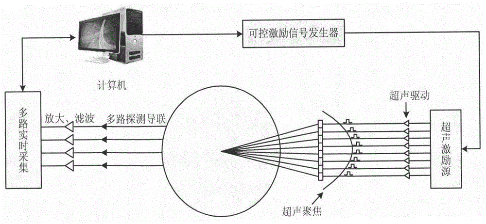
[0025] Such as figure 1 As shown, the biological tissue current density imaging method based on the acoustoelectric effect of the present invention comprises the following steps:

[0026] (1) Ultrasonic excitation source is used to generate ultrasonic waves, which are driven by ultrasonic waves and focused on the inside of biological tissues;

[0027] (2) Continuous positioning of ultrasonic waves focused on the inside of biological tissue, so as to complete the overall or partial scanning of biological tissue;

[0028] (3) Signal acquisition: Ultrasound is focused and positioned at a certain spatial position, and a high-frequency signal corresponding to the current density of the spatial position will be generated. The signal is collected by multiple detection leads and output after amplification and filtering. After ultrasonic focused scann...


PUM


Abstract

The invention discloses an imaging method of the biological tissue electric current density based on the acoustoelectric effect. The imaging method of the biological tissue electric current density based on the acoustoelectric effect comprises the following steps: (1) adopting an ultrasound excitation source to generate ultrasonic wave, driving and focusing the ultrasonic wave at the inner portion of biological tissue by the ultrasound; (2) continuously positioning the ultrasonic wave which is focused at the inner portion of the biological tissue, and finishing scanning of the whole or the local of the biological tissue; (3) collecting signals, focusing and positioning in a certain spatial position by the ultrasonic wave, generating a high frequency electrical signal which corresponds to the electric current density in the spatial position, and obtaining a set of signals of the whole or the local of the biological tissue; and (4) calculating and rebuilding the electric current density at the inner portion of the biological tissue according to the signals obtained after the scanning of the whole or the local of the biological tissue. The imaging method of the biological tissue electric current density based on the acoustoelectric effect is dynamic imaging, can intuitively reflect internal activities in the biological tissue, can obtain an electric current density image which is high in precision and resolution, and can facilitate medical diagnosis.

Description

technical field [0001] The invention relates to the field of biological tissue imaging, in particular to a biological tissue current density imaging method based on the acoustoelectric effect. Background technique [0002] Medical images are intuitive, vivid and informative, and play an important role in clinical diagnosis. With the advancement of medical imaging technology, medical imaging has developed from anatomical structure imaging to functional imaging, that is, medical images can not only display the morphological changes of the anatomical structure of the disease, but also reflect the functions of the body's organs and related physiological and biochemical changes, and can detect The abnormality of its physiological activities provides important information for disease diagnosis and promotes the progress and development of medicine. Taking the heart as an example, arrhythmia is a major clinical disease that endangers human health. Determining the location of the le...

Claims


Application Information

Patent Timeline
Patent Type & Authority Patents(China)
IPC IPC(8): A61B5/053A61B8/08
Inventor 徐文龙徐冰俏张宁宋兴辉
Owner CHINA JILIANG UNIV
Why Eureka
Try Eureka