A type Ⅱ adeno-associated virus carrying neuritin gene and its application in repairing optic nerve damage
A gene and virus technology, applied in the repair of optic nerve damage, in the field of adeno-associated virus type II, to improve survival and regeneration, enhance intrinsic regeneration ability, and promote optic nerve regeneration.
- Summary
- Abstract
- Description
- Claims
- Application Information
AI Technical Summary
Problems solved by technology
Method used
Image
Examples
Embodiment 1
[0044] 1. Construction of Neuritin gene type Ⅱ adeno-associated virus expression vector
[0045] (1) Type II adeno-associated virus expression vector and enzyme digestion of target gene
[0046] The pAOV-CAG-eGFP vector (purchased from Addgene) was used to carry out enzyme digestion with NotI and MluI, and the digested vector was prepared for use in vector construction. Using the cDNA clone of the CDS region of the NRN1 gene as a template, the NRN1 gene fragment was amplified by PCR, and the product was digested with NotI and MluI, and the vector and target gene DNA were recovered after identification by agarose gel electrophoresis.
[0047] (2) Ligation reaction
[0048] The digested type II adeno-associated virus vector and the PCR product of the NRN1 gene were ligated according to the reaction system in Table 1.
[0049] Table 1: Ligation reaction system
[0050]
[0051] After the above reagents were mixed, they were ligated at 4°C for 12 hours to prepare the cloning l...
Embodiment 2
[0066] 1. Culture and induction of RGC5 differentiation
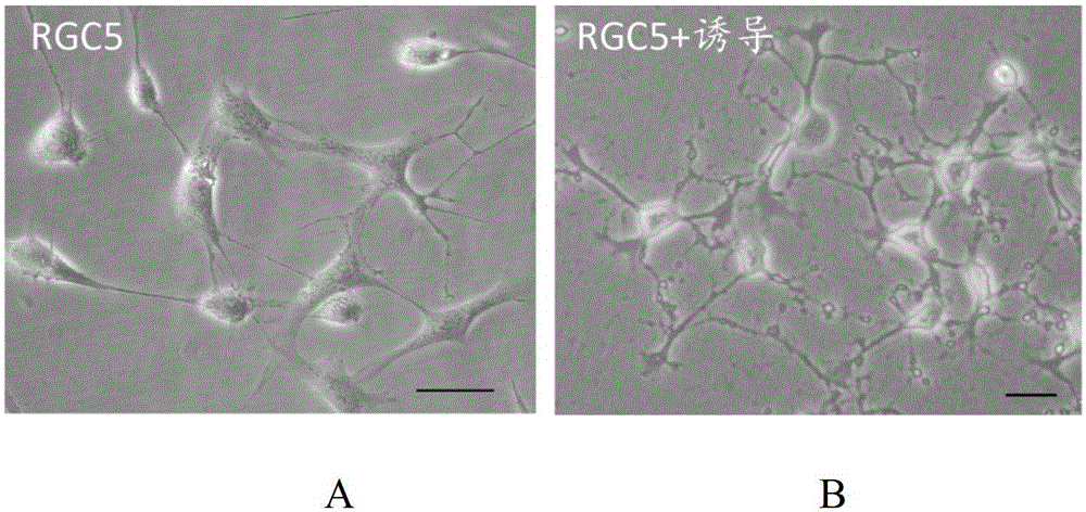
[0067] RGC5 is the retinal ganglion cell line of the one-day-old rat (see [20] KrishnamoorthyRR, AgarwalP, PrasannaG, etal.Characterizationofatransformedradretinalganglioncellline.BrainResMolBrainRes.2001;86(1-2):1-12.), immature , first induced by staurosporine to differentiate into mature neuron-like cells ( figure 1 ). RGC5 before induction was cultured in high-glucose DMEM medium and 5% serum in a place containing 5% CO 2 , in an incubator at 37°C, and the cells were passaged once a day. When used, 326nM staurosporine was used to induce for 24 hours.
[0068] 2. Overexpression of Neuritin gene and establishment of RGC5 injury model
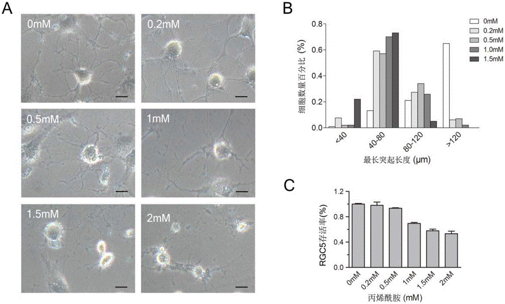
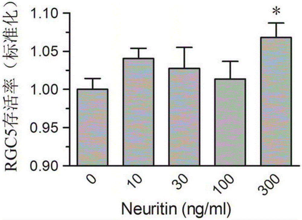
[0069] When RGC5 was cultured to 80% confluence, add the appropriate amount of type II adeno-associated virus and a certain amount of polybrene at a pre-measured concentration, and replace it with normal medium after 24 hours, observe the expression of RFP with a fluorescence micros...
Embodiment 3
[0081] 1. In vivo transfection of retinal ganglion cells with type Ⅱ adeno-associated virus and overexpression of Neuritin
[0082]Adeno-associated virus type II was injected into the vitreous body, and mice were perfused whole body with 4% paraformaldehyde one week later. Retinal slices were taken, and immunohistochemical β-Ⅲ-tubulin staining or cholera toxin B subunit (CTB) injection was used to co-localize the retinal ganglion cells to observe the transfection effect, the transfection efficiency is about 90% or more (such as Figure 5 shown).
[0083] Western blot and immunohistochemical methods were used to observe the overexpression and localization of Neuritin protein after Neuritin type Ⅱ adeno-associated virus transfection.
[0084] 2. Establishment of Optic Nerve Injury Model
[0085] The present invention adopts the optic nerve crush model, using Yasargil cerebral aneurysm clip on the optic nerve of adult male rats at about 1mm behind the eyeball to crush for 9 sec...
PUM
 Abstract
Description
Claims
Application Information
 - R&D
- Intellectual Property
- Life Sciences
- Materials
- Tech Scout
- Unparalleled Data Quality
- Higher Quality Content
- 60% Fewer Hallucinations
Browse by: Latest US Patents, China's latest patents, Technical Efficacy Thesaurus, Application Domain, Technology Topic, Popular Technical Reports.
© 2025 PatSnap. All rights reserved.Legal|Privacy policy|Modern Slavery Act Transparency Statement|Sitemap|About US| Contact US: help@patsnap.com


