Random positioning super-resolution microscopy method and device based on fluorescence emission suppression mechanism

A fluorescence emission and super-resolution technology, used in fluorescence/phosphorescence, material excitation analysis, etc., which can solve problems such as imperfection, damage to observed samples, and cumbersome sample processing.

Active Publication Date: 2016-01-20
CHINA JILIANG UNIV +1
View PDF11 Cites 0 Cited by
  • Summary
  • Abstract
  • Description
  • Claims
  • Application Information

AI Technical Summary

Problems solved by technology

The electron microscopic imaging technology developed in the 1930s and various non-optical probe scanning microscopic imaging technologies that emerged in the early 1980s have nanometer or even higher resolution capabilities, but they exist to varying degrees. The complex structure of the system, the harsh requirements of the imaging and detection environment, and the cumbersome sample processing are difficult, especially the important optical information of the sample (such as reflectivity, refractive index, polarization state, and spectrum, etc.) cannot be obtained, so it cannot completely replace optical microscopic imaging. status
Although the above two methods can achieve the purpose of super-resolution microscopy, they are not perfect. For example, the target switching and reading microscopy techniques need to apply strong suppression light to the sample (generally hundreds of megaW / cm 2 ); while random switch and readout microscopy requires pre-bleaching of fluorescent molecules in the observation area
Both of these situations are prone to irreversible damage to the observed sample

Method used


Image

  • Random positioning super-resolution microscopy method and device based on fluorescence emission suppression mechanism
  • Random positioning super-resolution microscopy method and device based on fluorescence emission suppression mechanism
  • Random positioning super-resolution microscopy method and device based on fluorescence emission suppression mechanism

Examples


Embodiment Construction

[0063] The present invention will be described in detail below in conjunction with the accompanying drawings, but the present invention is not limited thereto.

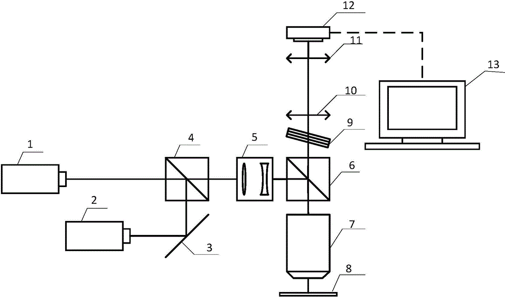
[0064] Such as figure 1 As shown, the randomly positioned super-resolution microscopy device based on the fluorescence emission suppression mechanism includes: a first laser light source 1, a second laser light source 2, a mirror 3, a first dichromatic mirror 4, a Kohler mirror group 5, a second two Color mirror 6, microscope objective lens 7, sample 8, optical filter 9, field lens 10, eyepiece 11, wide field photosensitive element 12 and computer 13.

[0065] In addition to the computer 13, the optical elements are arranged along the optical path direction, the first laser light source 1 and the first dichromatic mirror 4 are all located on the main axis optical path, and the second laser light source 2 converges with the first laser light source 1 on the first two through the reflector 3. On the color mirror 4 , th...


PUM

PropertyMeasurementUnit
poweraaaaaaaaaa
poweraaaaaaaaaa

Abstract

The invention discloses a random positioning super-resolution microscopy method and device based on a fluorescence-emission kill mechanism. The method includes the following steps: coaxial and common-path exciting light and restraining light are simultaneously focused on a sample; the position on the sample, with the stimulated fluorescence-emission characteristic, randomly emits fluorescent light; fluorescent signals are collected to generated a sparse fluorescent distribution image; diffraction spots are positioned in a unimolecule manner, and the final product is obtained after different fluorescent light positioning images are synthesized. The device includes a first laser light source, a second laser light source, a reflector, a first dichroic mirror, a kohler mirror group, a second dichroic mirror, a microobjective, a sample, an optical filter, a field lens, an ocular, a wide field sensing element and a computer. The invention has the advantages that the resolution ratio and fineness are high, 20 nm crosswise super-resolution images can be obtained; the structure is simple, and the cost is low; irreversible damage of intensive laser or fluorescence photobleaching to samples is reduced, and repeating utilization ratio of samples is increased; the function extensibility is strong.

Description

technical field [0001] The invention relates to the field of microscopic observation methods and equipment for biological samples, in particular to a random positioning super-resolution microscopy method and device based on a fluorescence emission suppression mechanism. Background technique [0002] Nanotechnology and biotechnology are the fastest-growing and hottest scientific fields in the 21st century. Nanotechnology has a wide range of applications, including imaging, measurement, processing, manipulation, etc. within the scale of 1-100nm. Many important organisms such as glucose, antibodies, viruses, etc. are in this scale range, and the need to study these tiny objects has driven the development of high-resolution microscopy. In turn, the development of super-resolution microscopy imaging technology has also promoted the progress of the entire life sciences. A major advantage of light microscopy over other microscopy techniques is the ability to study living cells in...

Claims


Application Information

Patent Timeline
Patent Type & Authority Patents(China)
IPC IPC(8): G01N21/64
Inventor 李旸晖金尚忠袁琨金杯洲
Owner CHINA JILIANG UNIV
Why Eureka
Try Eureka