Multi-modal homomorphic isochronic medical image imaging system and method

An imaging system and imaging method technology, applied in the field of biomedical imaging, can solve problems such as low sensitivity, inability to detect multiple molecules at the same time, and inability to meet the urgent needs of life science research, and achieve the effect of accurate image results

Active Publication Date: 2014-05-28
北京锐视康科技发展有限公司
View PDF5 Cites 17 Cited by
  • Summary
  • Abstract
  • Description
  • Claims
  • Application Information

AI Technical Summary

Problems solved by technology

[0003] Any molecular imaging with a single modality has its own insurmountable defects, which cannot meet the urgent needs of life science research - to fully and accurately explain the life process
For example, although Positron Emission Tomography (Positron Emission Tomography) PET has high detection sensitivity, it cannot detect multiple molecules at the same time because the radiation photon energy is fixed at 511keV; Single Photon Emission Computed Tomography (Single Photon Emission Computed Tomography) SPECT Although there is no need for expensive cyclotrons to prepare nuclide drugs, it is difficult to find SPECT radiotracers involved in metabolism, and its sensitivity is 1 to 2 orders of magnitude lower than that of PET
At present, the three modalities of X-ray CT, PET, and SPECT have been combined using hardware methods, but the imaging of the three modalities is not homogeneous and isochronous data acquisition
The imaging time of nuclear medicine imaging methods such as PET and SPECT is generally tens of minutes, so multi-modal dynamic acquisition with high isochronism requirements is often unsatisfactory
For the field of molecular medical imaging, the multi-modal imaging devices in the prior art place each imaging modality in a different position, fail to share the same scanning area, and can only use time-sharing scanning, which cannot Achieve X-ray CT / PET / SPECT three-modal isochronous scanning imaging

Method used


Image

  • Multi-modal homomorphic isochronic medical image imaging system and method
  • Multi-modal homomorphic isochronic medical image imaging system and method
  • Multi-modal homomorphic isochronic medical image imaging system and method

Examples


Embodiment Construction

[0029] Below in conjunction with accompanying drawing, the present invention will be further described by examples.

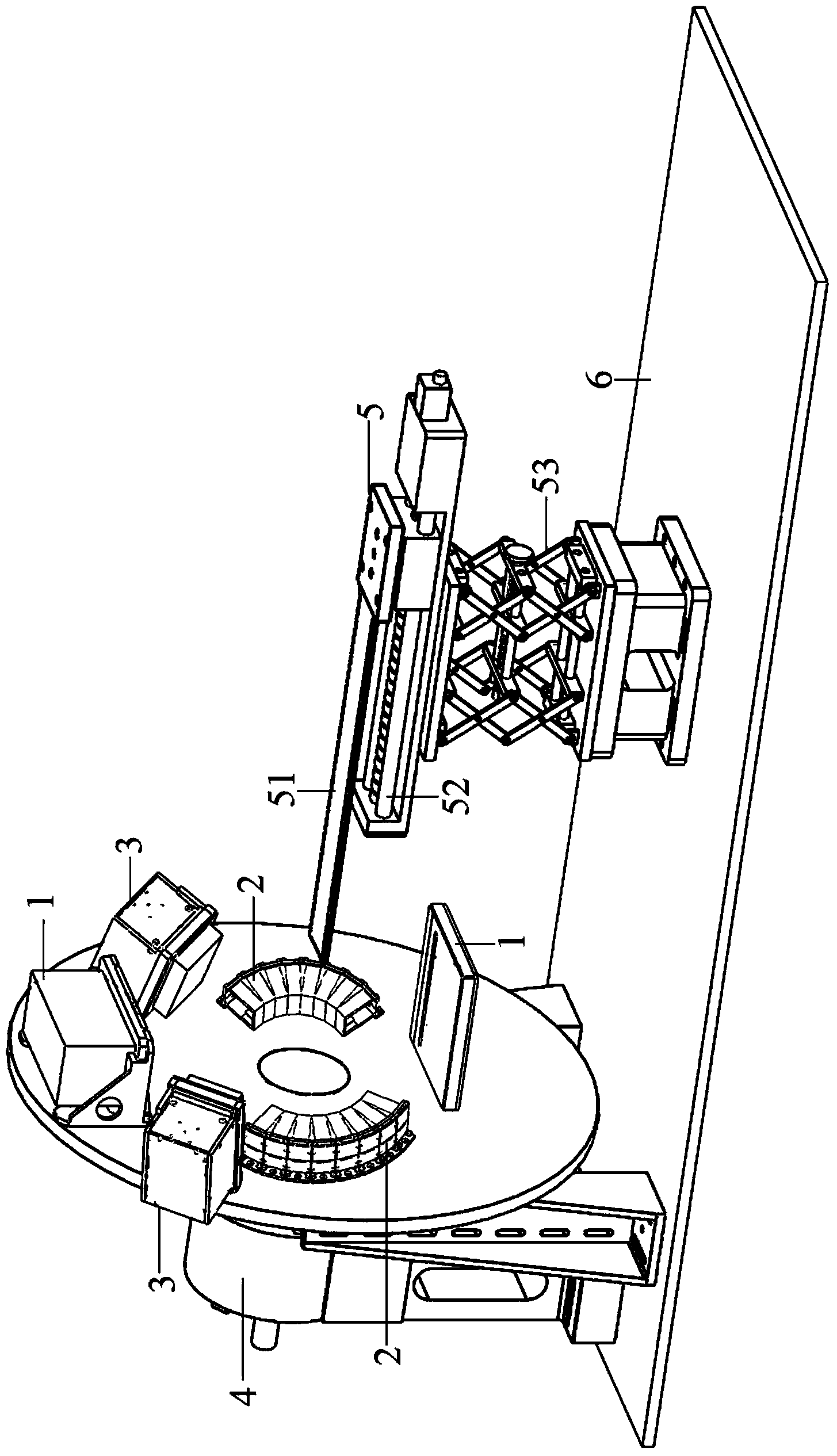
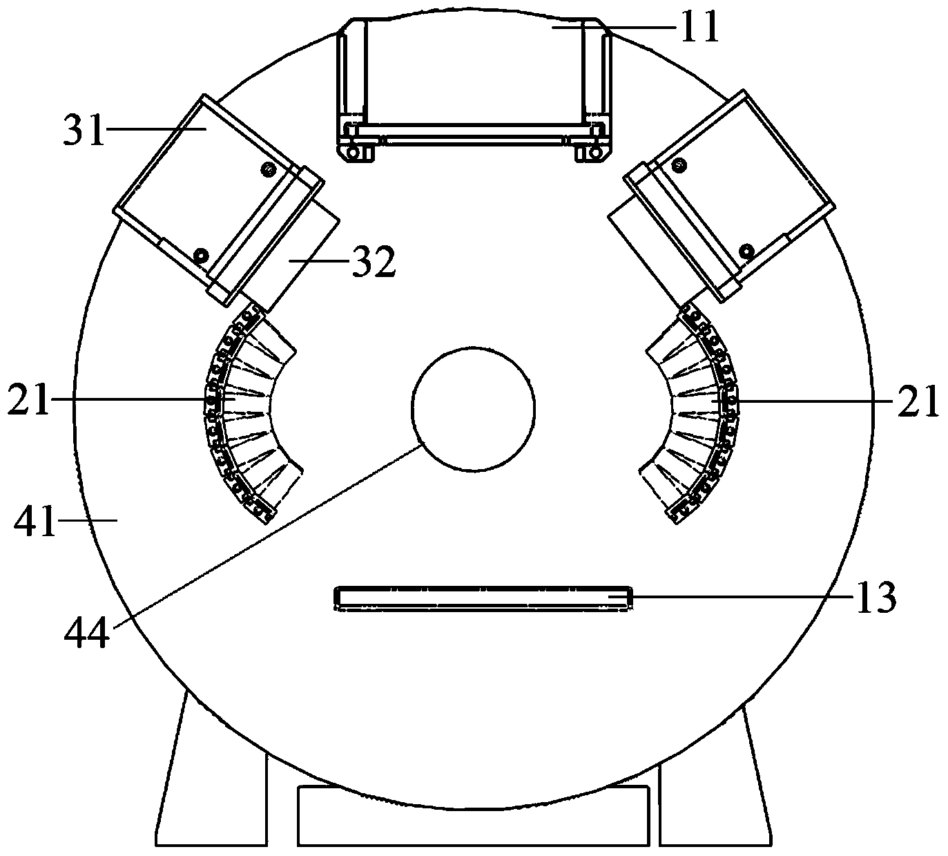
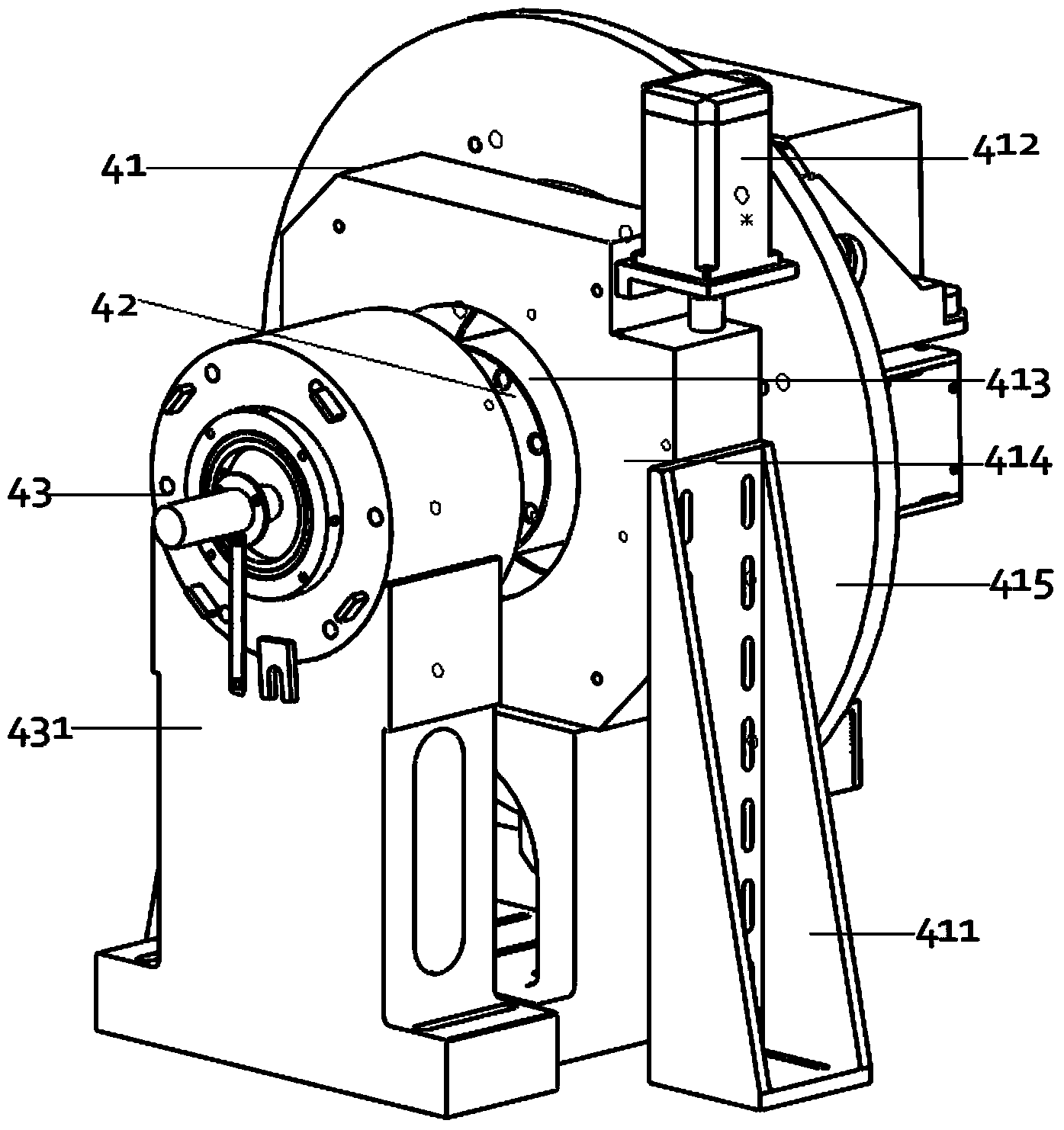
[0030] like figure 1 As shown, in this embodiment, the multimodal homomorphic isochronous medical imaging system includes: X-ray computed tomography X-ray CT device 1, positron emission tomography PET device 2, single photon emission tomography SPECT device 3. Rotating device 4, scanning bed device 5, and data acquisition system and computer; wherein, X-ray CT device 1, PET device 2 and SPECT device 3 are installed on the same rotating device 4 to form a multimodal imaging system, Installed on one end of the base 6; the scanning bed device 5 is installed on the other end of the base, and the scanning tracks of each imaging device are in the same horizontal plane; each imaging device is sampled and saved to the computer by the data acquisition system through the data line; each imaging device shares a The scanning table assembly and the same scanning area.

[...


PUM


Abstract

The invention discloses a multi-modal homomorphic isochronic medical image imaging system and a multi-modal homomorphic isochronic medical image imaging method. The imaging system comprises an X-ray computed tomography (X-ray CT) device, a positron emission tomography (PET) device, a single photon emission computed tomography (SPECT) device, a rotating device, a scanning table device, a data acquisition system and a computer, wherein the X-ray CT device, the PET device and the SPECT device are arranged on the same rotating device, and share one scanning table device and the same scanning area to form the multi-modal imaging system. The same sampling angle is subjected to time-share scanning in three modes and is rotated by one circle, multi-modal homomorphic isochronic imaging is realized, different imaging equipment complements advantages of one another, the acquired image results are more accurate and reliable, multi-modal dynamic acquisition in real sense is realized.

Description

technical field [0001] The invention relates to the field of biomedical imaging, in particular to a multimodal homomorphic isochronous medical imaging system and an imaging method thereof. Background technique [0002] Medical imaging technology has always been an important means of diagnosing diseases, and it is also the earliest engineering means used by people for medical diagnosis. In 1895, German scientist Wilhelm Conrad An unknown ray that can penetrate matter was discovered while experimenting with cathode ray tubes, Roentgen Name it "X-Ray". Mrs. Roentgen's hand bone X-ray angiography also created a radiology using X-rays for medical diagnosis - X-ray angiography, and also created a new era of the combination of engineering technology and medicine. Computed Tomography began in the 1860s or earlier, and it was not until the 1980s that the systematic study of tomographic image fusion began. Following the early flat-panel image fusion, until the 1990s, the two bas...

Claims


Application Information

Patent Timeline
Patent Type & Authority Applications(China)
IPC IPC(8): A61B6/03
Inventor 任秋实卢闫晔杨昆
Owner 北京锐视康科技发展有限公司
Why Eureka
Try Eureka