
Medical device and methods for blood vessel compression

A technology for medical equipment and vascular compression, applied in mechanical equipment, applications, tourniquets, etc., can solve the problems of complex structure and cannot provide sufficient pressure control, and achieve the effect of simple structure, simple application, and ensuring pressure control.

Active Publication Date: 2014-06-25
SCHOOL OF CARDIOLOGY
View PDF8 Cites 3 Cited by
  • Summary
  • Abstract
  • Description
  • Claims
  • Application Information

AI Technical Summary

Problems solved by technology

[0005] Radial compression devices known in the prior art do not provide an adequate level of pressure control when they are applied, or are of very complex construction and require pressure control devices or additional equipment (such as pneumatic syringes) to be fully functional

Method used


Image

  • Medical device and methods for blood vessel compression
  • Medical device and methods for blood vessel compression
  • Medical device and methods for blood vessel compression

Examples


Embodiment Construction

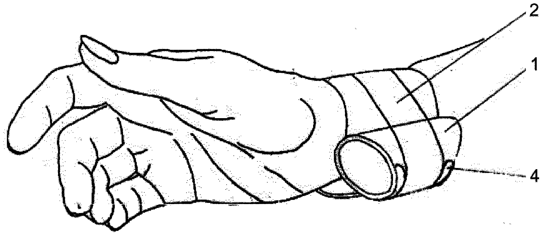
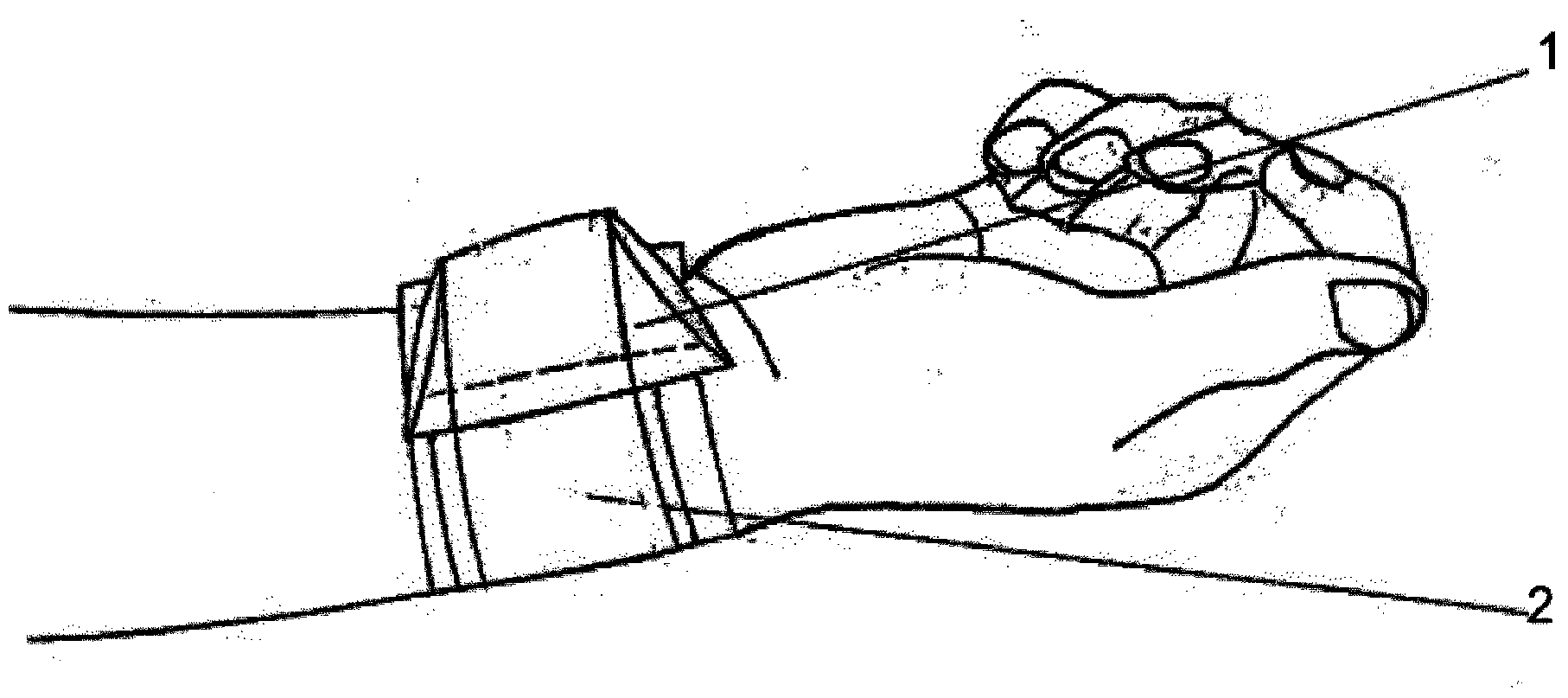
[0069] in figure 1 A first preferred embodiment of the medical device of the present invention is shown in, which is fastened to the forearm near the wrist, above the radial artery ra and opposite to the ulnar artery ua. The device includes a main body 1, which is made of a transparent material and is shaped into a hollow cylinder with beveled ends. A holding element 2 in the form of a transparent strip is attached to the outer surface of the main body 1. The main body 1 is equipped with two guiding devices 4 which guide the strap into an appropriate position above the main body 1 when the device is applied to the patient's limbs. The strap is wrapped around the patient's limb, and the main body 1 is fastened in place. The position of the strap ensures that the required compression force is applied to the patient's limb at the contact site.

[0070] in image 3 , Figure 4 , Figure 5 with Image 6 A more detailed view of the first preferred embodiment of the device is present...


PUM


Abstract

The invention relates to a medical device and method for applying pressure onto a patient's limb, especially at a blood vessel or a wound site, in order to achieve local hemostasis. The device comprises a body (1) for blood vessel compression, holding element (2) for attaching the body (1) to a patient's limb, a fastening means (3) for holding the device in a desirable position, wherein the body (1) has a first compression area (1a) which is situated at the outer surface of the body (1), at least one second compression area (1b) through which the body (1) is pressed with a holding element (2), a third compression area (1c) for compression control during application of the device, and wherein the holding element (2) is guided over the first compression area (1a), when the device is attached to the patient's limb.

Description

Technical field [0001] The present invention generally relates to medical devices and methods for applying pressure to a patient's limbs, especially to blood vessels or wound sites, to achieve local hemostasis. More specifically, in invasive cardiology and radiology, blood vessels are removed after cardiac catheterization (coronary angiography) or other operations and interventions that require vascular access ports for endovascular surgery During the vascular sheath, the device of the present invention is used. This device allows obstruction of the outflow of blood accompanying the removal of the vascular sheath. Background technique [0002] Currently known technical solutions aimed at achieving proper hemostasis usually require performing a series of consecutive actions one after another, usually by at least two people (a doctor and a nurse in most cases) working together. As part of the action, a strap is placed around the patient’s arm, then the strap is fastened to the wr...

Claims


Application Information

Patent Timeline
Patent Type & Authority Applications(China)
IPC IPC(8): A61B17/132
CPCA61B17/1325A61B2017/00893A61B2017/00902A61B2017/12004A61B2019/464A61B2017/00889A61B2090/064F04C2270/0421A61B17/12A61B17/132
Inventor M·克鲁克
Owner SCHOOL OF CARDIOLOGY
Learn More