Preparation method of cell slide
A cell and slide technology, applied in the field of cell slide preparation, can solve problems such as difficult quality control, inability to stack and overlap cells, growth density of slides, etc., and achieve the effect of reliable operation process
- Summary
- Abstract
- Description
- Claims
- Application Information
AI Technical Summary
Problems solved by technology
Method used
Image
Examples
Embodiment 1
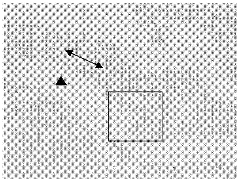
[0057] First, gently place 12 wet sterile slides into the bottom of a 12-well culture plate with tweezers. Then the cells in the logarithmic growth phase were taken, digested with preheated 0.25% trypsin, the cells were blown into a single cell suspension, and the cell density was adjusted to 1.25×10 5 About / ml, use a 1ml pipette gun to draw 1ml of the cell suspension, and vertically and quickly dispense the cell suspension at a distance of about 3cm from the bottom of the 12-well plate. 2 Cultivate in the incubator for 24h. After the cells adhered to the wall the next day, discard the culture medium, wash the cells once with PBS, and add serum-free DMEM culture medium, 1ml per well. Place the 12-well plate at 37°C, 5% CO 2 After continuing to culture in the incubator for 24 hours, take it out of the incubator, discard the supernatant, wash the cells with pre-cooled PBS for 3 times, add 1ml of 4% paraformaldehyde to fix for 30min, discard the fixative and then use the pre-c...
Embodiment 2
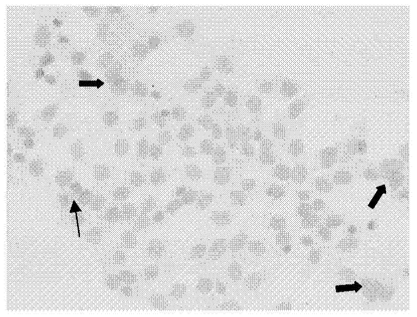
[0061] Use tweezers to lightly put the wet sterile slide into the bottom of the 12-well plate, then take the cells in the logarithmic growth phase, digest them with preheated 0.25% trypsin, blow the cells into a single-cell suspension, and separate the cells Density adjusted to 1.5×10 5 About / ml, use a 1ml pipette gun to draw 1ml of cell suspension, and vertically and quickly pipette the cell suspension at a distance of about 4cm from the bottom of the 12-well plate. 2 Cultivate in the incubator for 24h. After the cells adhere to the wall the next day, discard the culture medium, wash the cells once with PBS buffer, and add serum-free DMEM culture medium, 1ml per well. Place the 12-well plate at 37°C, 5% CO 2 After continuing to culture in the incubator for 24 hours, take it out of the incubator, discard the supernatant, wash the cells with pre-cooled PBS for 3 times, add 1ml of 4% paraformaldehyde for fixation for 40min, discard the fixative and then use the pre-cooled Wa...
Embodiment 3
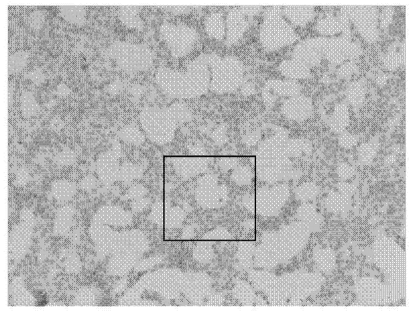
[0073] Use tweezers to lightly put the wet sterile slide into the bottom of the 12-well plate, then take the cells in the logarithmic growth phase, digest them with preheated 0.25% trypsin, blow the cells into a single-cell suspension, and separate the cells Density adjusted to 2.0×10 5 About / ml, use a 1ml pipette gun to draw 1ml of cell suspension, and vertically and quickly pipette the cell suspension at a distance of about 5cm from the bottom of the 12-well plate. 2 Cultivate in the incubator for 24h. After the cells adhered to the wall the next day, discard the culture medium, wash the cells once with PBS, and add serum-free DMEM culture medium, 1ml per well. Place the 12-well plate at 37°C, 5% CO 2 After continuing to culture in the incubator for 24 hours, take it out of the incubator, discard the supernatant, wash the cells with pre-cooled PBS for 3 times, add 1ml of 4% paraformaldehyde to fix for 50min, discard the fixative and then use the pre-cooled Wash with cold...
PUM
| Property | Measurement | Unit |
|---|---|---|
| Cell density | aaaaa | aaaaa |
Abstract
Description
Claims
Application Information
 - R&D
- Intellectual Property
- Life Sciences
- Materials
- Tech Scout
- Unparalleled Data Quality
- Higher Quality Content
- 60% Fewer Hallucinations
Browse by: Latest US Patents, China's latest patents, Technical Efficacy Thesaurus, Application Domain, Technology Topic, Popular Technical Reports.
© 2025 PatSnap. All rights reserved.Legal|Privacy policy|Modern Slavery Act Transparency Statement|Sitemap|About US| Contact US: help@patsnap.com


