
A microfluidic chip and a research method for cell chemotaxis

A microfluidic chip and cell technology, applied in the field of biomedical research, can solve the problems that do not have the characteristics of the closed space of microvessels, cannot reproduce the chemotaxis process of cells in microvessels, and cannot be used to study cell biological processes, etc., and has achieved wide application foreground effect

Active Publication Date: 2017-07-28
DALIAN MEDICAL UNIVERSITY
View PDF4 Cites 0 Cited by
  • Summary
  • Abstract
  • Description
  • Claims
  • Application Information

AI Technical Summary

Problems solved by technology

However, the Transwell chamber is a relatively large open space, which does not have the closed space characteristics of microvessels in the body; and because the cells in the chamber are in a static culture state, it is impossible to reproduce the chemotaxis process of cells in the microvessel under the flow state
Therefore, the Transwell chamber cannot be used to study the biological process of cells moving through the microvascular wall for directional chemotaxis under flow conditions.

Method used


Image

  • A microfluidic chip and a research method for cell chemotaxis
  • A microfluidic chip and a research method for cell chemotaxis
  • A microfluidic chip and a research method for cell chemotaxis

Examples


Embodiment 1

[0028] The microfluidic chip used was designed and manufactured by our laboratory.

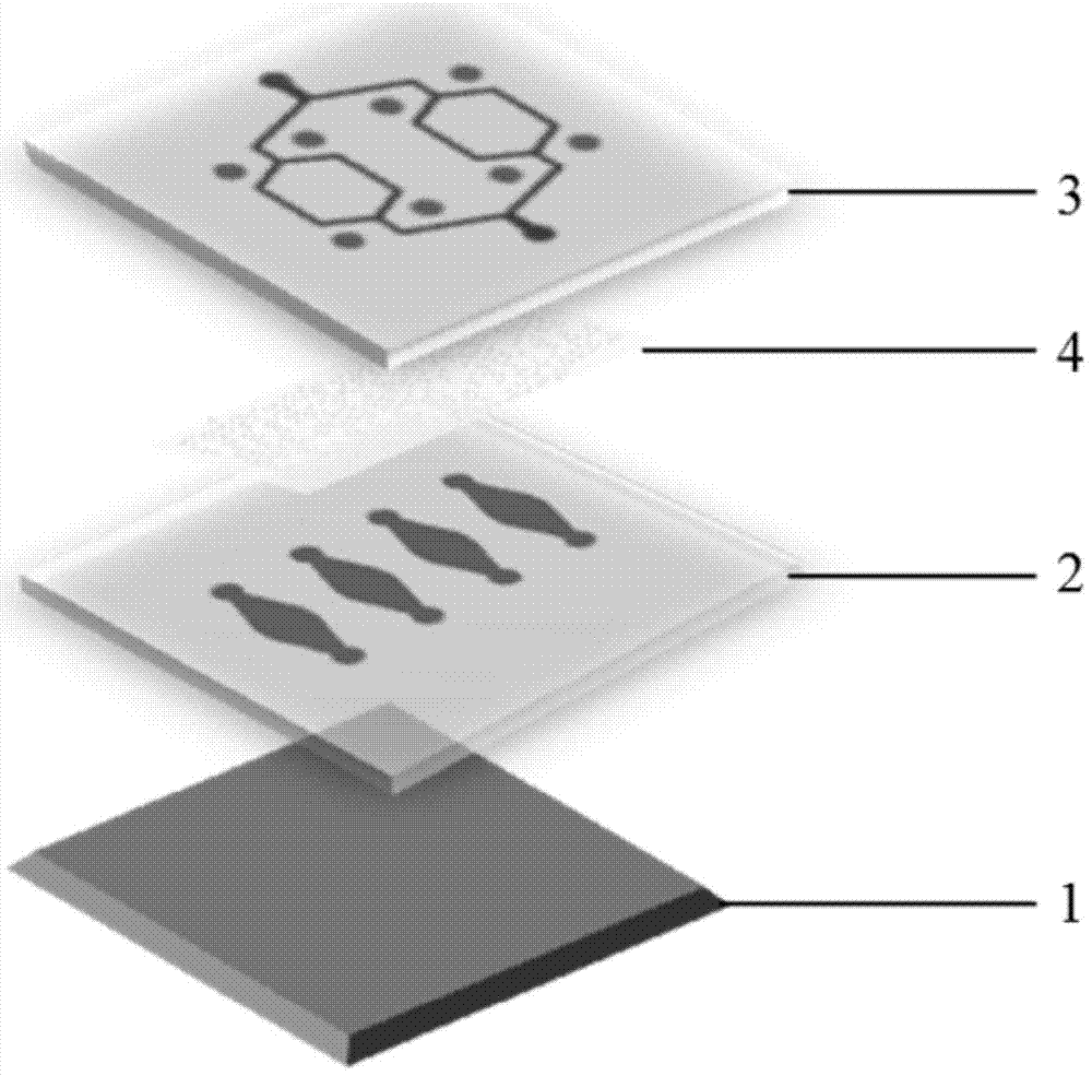
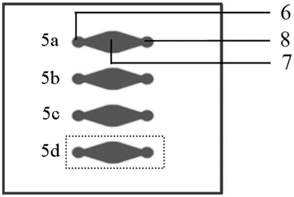
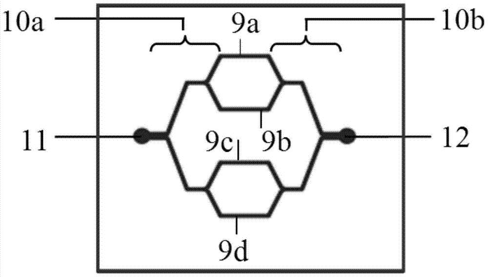
[0029] Such as Figure 1-3 As shown, the microfluidic chip of the present invention is composed of a glass substrate 1, a first membrane 2, a second membrane 3 and a porous membrane 4, wherein the material of the first membrane 2 and the second membrane 3 is polydimethylform base siloxane, and the porous membrane 4 is a polycarbonate membrane. There are four parallel and independent culture units (5a, 5b, 5c, 5d) on the first diaphragm 2, and each culture unit (5a, 5b, 5c, 5d) has an independent liquid inlet-6 and liquid outlet respectively Port one 8, a culture pool 7 is arranged between the liquid inlet one 6 and the liquid outlet one 8; there are four parallel microchannels (9a, 9b, 9c, 9d) on the second diaphragm 3, one of the microchannels One side is collected in the liquid inlet 2 11 through a group of branched connection channels 10a on one floor, and the other side is collected in t...

Embodiment 2

[0033] The chip described in Example 1 was used for research: FITC-Dextran (molecular weight is about 12KD) was added into the culture pool 7 through the liquid inlet 6, and as time went on, FITC-Dextran diffused through the porous membrane 4 and vascular endothelial cells into the microchannel of the second diaphragm 3 ( Image 6 ).

Embodiment 3

[0035]The chip described in Example 1 is used for research: through the liquid inlet 6, add the concentration of 0ng / ml, 25ng / ml, 50ng / ml, and 100ng / ml respectively in the four culture pools 7 of the first membrane 2. Chemokine CXCL12, incubated for 2 hours. Add the red fluorescent probe-labeled tumor cell line (ACC-M) into the microchannel through the liquid inlet 2 11 . Drive the red fluorescent probe-labeled tumor cell line (ACC-M) through the syringe pump 13 connected to the liquid inlet 2 11 to make it flow in the microchannel of the second diaphragm 3, stop after 30 minutes, and use phosphate After gently rinsing with buffer, take pictures under a fluorescent microscope to record the adhesion of ACC-M cells to vascular endothelial cells ( Figure 7 ).


PUM

PropertyMeasurementUnit
depthaaaaaaaaaa

Abstract

The aim of the invention is to provide a micro-fluidic chip and a cell chemotaxis movement research method. The micro-fluidic chip is characterized in that the micro-fluidic chip comprises a glass substrate, a first diaphragm and a second diaphragm; the first diaphragm is provided with culturing units; the second diaphragm is provided with micro-channels; a porous membrane exists between the culturing units of the first diaphragm and the micro-channels of the second diaphragm; the number of the culturing units of the first diaphragm is same to the number of the micro-channels of the second; and the micro-channels on the second diaphragm are respectively located above the culturing units of the first diaphragm. The micro-fluidic chip can be used in the research of the selective chemotaxis movement condition of cells in the micro-channels when the cells flow through the culturing units. The micro-fluidic chip likes a bionic model characterized in that a branching capilary goes through organs; and compared with the traditional cell chemotaxis research methods, the research method is a new method for researching the selective chemotaxis movement of the cells in the flowing process, and has important biomedical research values and economic values.

Description

technical field [0001] The invention relates to the application of microfluidic chip technology to the field of biomedical research. The invention particularly provides a microfluidic chip and a research method for studying cell chemotaxis movement. Background technique [0002] Chemotaxis refers to the process in which cells move toward stimuli under the action of chemical concentration gradients. Chemotaxis plays an important role in the development of tissues and organs, wound healing and tumor metastasis. For example, injured tissues secrete chemokines to attract leukocytes to the infection site, remove necrotic tissue, and promote wound healing; organs such as the lung, liver, and bone are It secretes a large number of chemokines and becomes the most common target organ of tumor metastasis. [0003] At present, the commonly used in vitro model for studying cell chemotaxis is the Transwell chamber, which is mainly used to study the chemotaxis process of cells in a stat...

Claims


Application Information

Patent Timeline
Patent Type & Authority Patents(China)
IPC IPC(8): C12M1/34C12Q1/02
CPCB01L3/5027B01L2200/0636B01L2200/0647B01L2300/0887G01N33/5029
Inventor 刘婷姣孔晶
Owner DALIAN MEDICAL UNIVERSITY
Learn More