Method for establishing goose embryo epithelial cell line and established goose embryo epithelial cell line
A technology for epithelial cells and goose embryos, which is applied in the field of established goose embryo epithelial cell lines to achieve the effects of high purity, high success rate and less restriction
- Summary
- Abstract
- Description
- Claims
- Application Information
AI Technical Summary
Problems solved by technology
Method used
Image
Examples
Embodiment 1
[0078] Example 1 Establishment of Goose Embryo Epithelial Cell Line
[0079] Establish the goose embryo epithelial cell line of the present invention according to the following steps:
[0080] (1) Aseptically take 11-13-day-old goose embryo tissue, cut off the head, limbs and internal organs with sterilized scissors, put it into a 10mL glass beaker, add 10mL PBS, and wash for 5-10 minutes; discard; After removing the PBS, add 10-20 mL of gentamicin solution prepared with PBS with a concentration of 20% to 70% by mass for 5-10 minutes;
[0081] (2) Take out the duck embryo tissue and cut it into 0.5~1.5mm3 tissue blocks. Place the tissue blocks flat on the bottom of the 6-well culture plate, one piece for each hole, and add 2~3mL to the culture hole containing 10% fetal calf by volume. Serum, 1%-2% goose serum and 0.2%-1% goose embryo allantoic fluid in DMEM culture medium, cultured at 37℃ and 5% CO2 until the tissue block is completely attached to the wall, then add the above DMEM m...
Embodiment 2
[0092] Example 2 Analysis of biological characteristics of goose embryo epithelial cell line
[0093] 1. Morphological observation
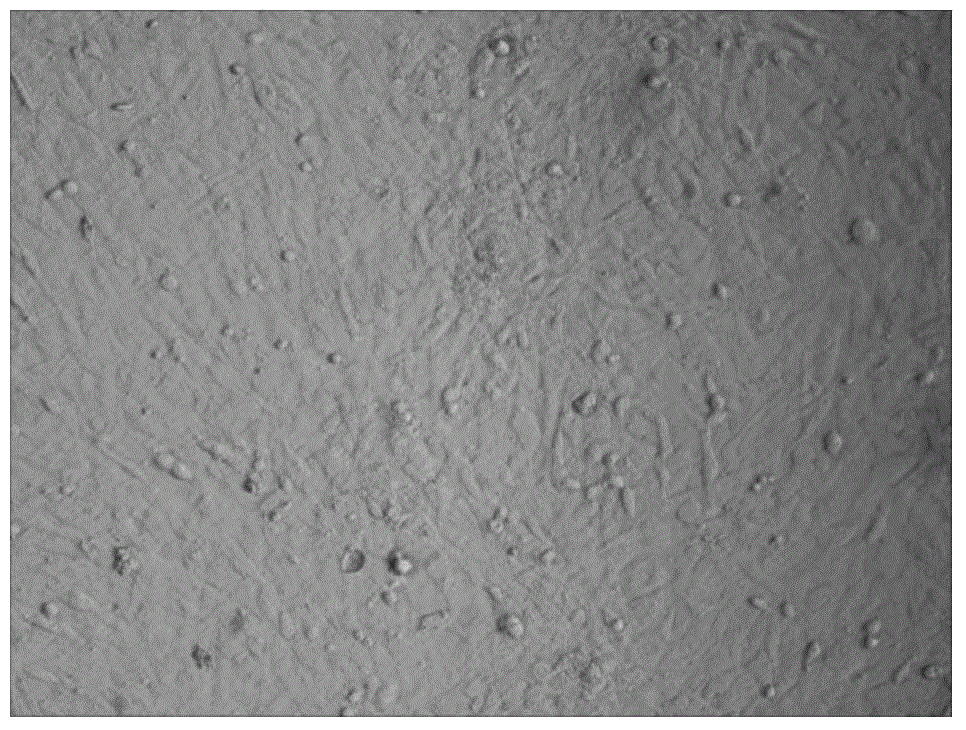
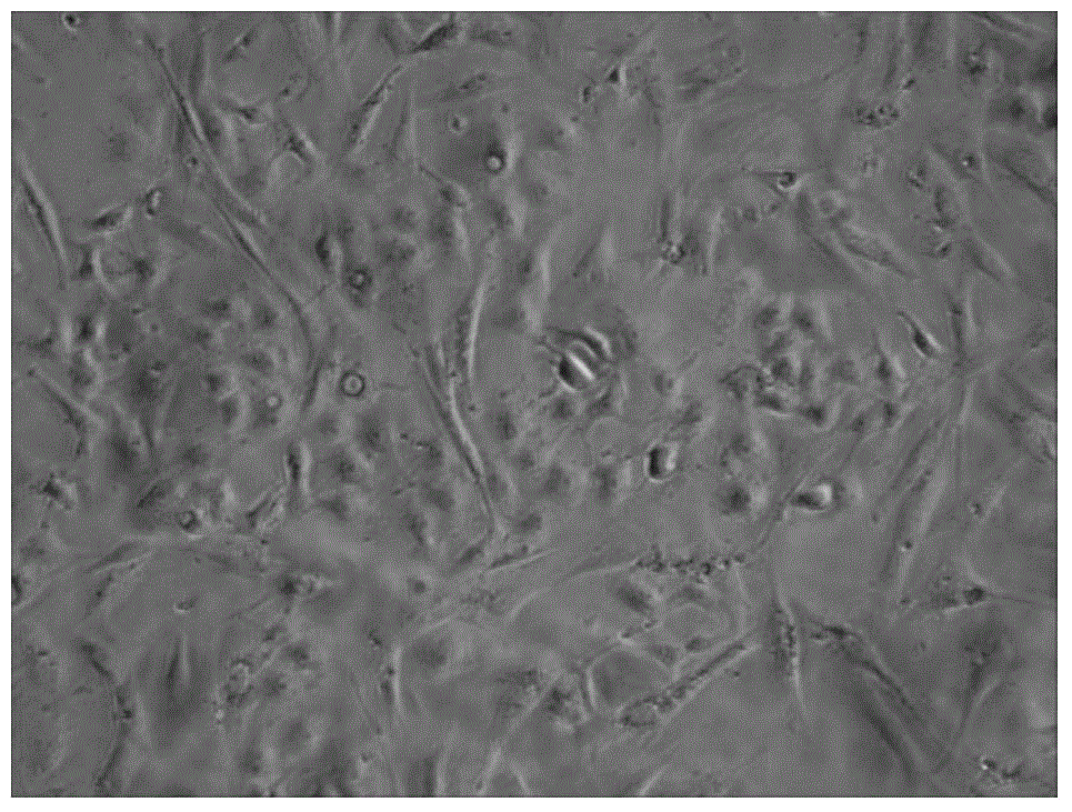
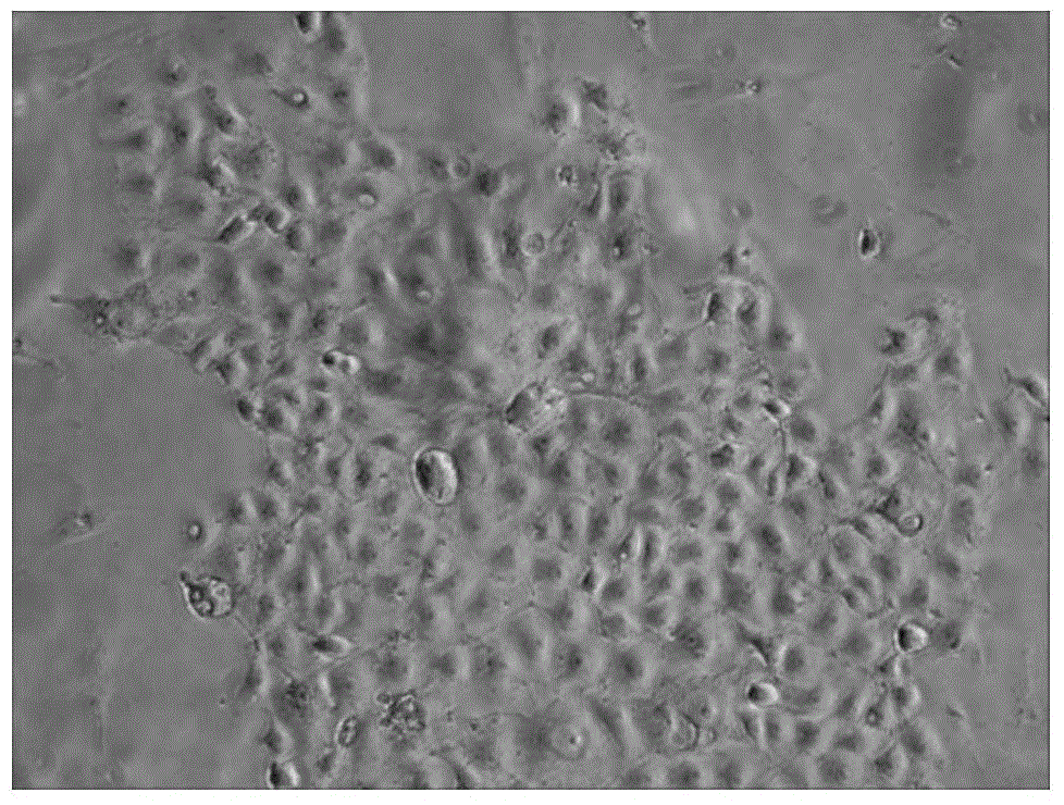
[0094] Observed by an inverted microscope, the established goose embryo epithelial cell line F30 generation ( Figure 4 ) And F50 generation ( Figure 5 ) And primary cells ( figure 1 ) Compared to the morphology, the primary cells contain a variety of heterogeneous cells, most of the cells are long spindle-shaped, a few are polygonal and oval, and the established epithelial cell line is obtained from a single cell, its purity Above 99.9%, they are all epithelioid cells. HE staining also shows that the cells are polygonal or short spindle-shaped epithelioid cells ( Image 6 ), with a round nucleus, cell growth and division ability is very strong, population doubling time is only 17.1h.
[0095] 2. Growth curve determination
[0096] Take the 30 generations and 50 generations of the cells to be tested in a good growth state, and when they are close to con...
Embodiment 3
[0102] Example 3 Verification of the sensitivity of goose embryo epithelial cell line to Muscovy Duck Parvovirus
[0103] 1. Method
[0104] Take 50th passage cells of goose embryo epithelial cell line, aspirate and discard the culture medium when it grows to 80% monolayer, wash twice with D-Hanks medium, inoculate Muscovy parvovirus, absorb at 37°C for 1 hour, discard the virus solution And supplemented with DMEM maintenance solution (containing 1% newborn calf serum) culture, and set up a blank control (goose embryo epithelial cell line not inoculated with Muscovy Duck Parvovirus). The same method is used to inoculate gosling plague virus, type I duck hepatitis virus, and new duck hepatitis virus.
[0105] 2. Results
[0106] It was observed that the goose embryo epithelial cells were sensitive to Muscovy Duck Parvovirus, and the infected cells within 96 hours all produced unique cytopathic changes, see Picture 11 ; While the control cell morphology is normal, see Picture 10 .
PUM
 Abstract
Description
Claims
Application Information
 - R&D
- Intellectual Property
- Life Sciences
- Materials
- Tech Scout
- Unparalleled Data Quality
- Higher Quality Content
- 60% Fewer Hallucinations
Browse by: Latest US Patents, China's latest patents, Technical Efficacy Thesaurus, Application Domain, Technology Topic, Popular Technical Reports.
© 2025 PatSnap. All rights reserved.Legal|Privacy policy|Modern Slavery Act Transparency Statement|Sitemap|About US| Contact US: help@patsnap.com


