PCT (procalcitonin) and CRP quantitative joint inspection chromatography test strip and preparation method thereof

An immunochromatographic test strip and detection line technology, applied in the field of medical testing, can solve the problems of lack of quantitative joint inspection of PCT and CRP, low sensitivity, immunotransmission turbidimetric methodology and clinical application need further verification, etc. Reduce the risk of errors, reduce costs, and reduce usage effectiveness

Active Publication Date: 2015-01-28
GUANGZHOU WONDFO BIOTECH
View PDF12 Cites 26 Cited by
  • Summary
  • Abstract
  • Description
  • Claims
  • Application Information

AI Technical Summary

Problems solved by technology

But the sensitivity is not high
[0012] 4. Transmission immunoturbidimetry - this method is simple, fast, automatic and suitable for batch detection, but the me

Method used


Image

  • PCT (procalcitonin) and CRP quantitative joint inspection chromatography test strip and preparation method thereof
  • PCT (procalcitonin) and CRP quantitative joint inspection chromatography test strip and preparation method thereof
  • PCT (procalcitonin) and CRP quantitative joint inspection chromatography test strip and preparation method thereof

Examples


Embodiment 1

[0057] Example 1 PCT and CRP quantitative combined detection immunochromatographic test strip and preparation method thereof

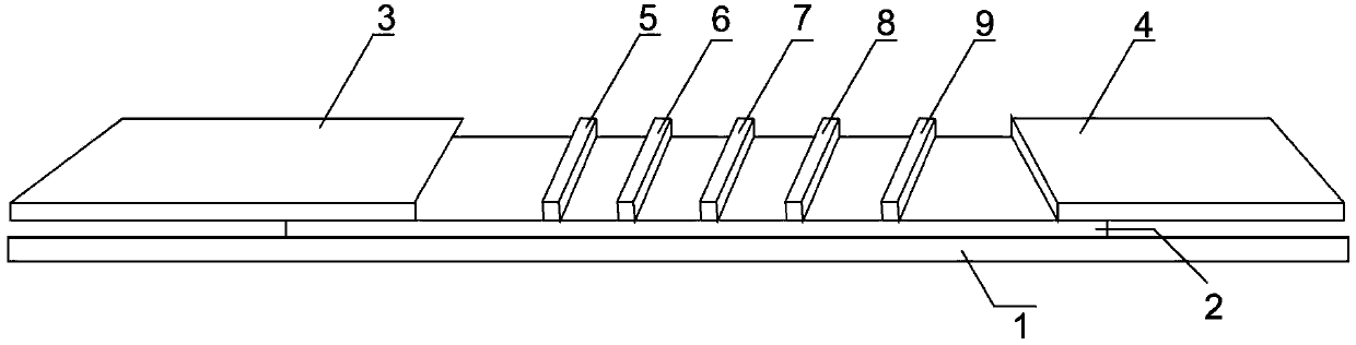
[0058] Such as figure 1 Shown is a structural schematic diagram of the PCT and CRP quantitative combined detection immunochromatographic test strip of the present invention. A PCT and CRP quantitative combined detection immunochromatographic test strip, comprising a substrate 1 and a coating film 2 arranged on the substrate 1, the coating film 2 is provided with a sample pad 3, a coating line area and Absorbent paper 4, the coated line area includes the fluorescent latex coated with CRP monoclonal antibody, PCT monoclonal antibody and goat antibody, which are arranged in parallel in turn near the 3rd end of the sample pad to the 4th end of the absorbent paper and are spaced apart from each other. The labeling line 5 of the chicken IgY antibody, the quality control line 6 coated with the chicken IgY antibody, the CRP detection line 7 coated with anothe...

Embodiment 2

[0074] Example 2 PCT and CRP quantitative combined detection immunochromatographic test strip and preparation method thereof

[0075] The structure of the test strip of this embodiment is the same as that of the test strip of Example 1. The difference is that in this embodiment, the coating membrane 2 is a nitrocellulose membrane with a climbing speed of 110 s / 4cm.

[0076] The preparation method of the PCT and CRP quantitative joint detection immunochromatography test strip of this embodiment is as follows, comprising the following steps:

[0077] 1. Covalent Activation of Fluorescent Latex

[0078] With embodiment 1.

[0079] 2. Preparation of Fluorescent Latex Microparticle-labeled Proteins

[0080] With embodiment 1.

[0081] 3. Preparation of Nitrocellulose Coated Membrane

[0082] a. Preparation of marker lines

[0083] Dilute the fluorescent latex labeled with CRP monoclonal antibody, the fluorescent latex labeled with PCT monoclonal antibody and the fluorescent l...

Embodiment 3

[0092] Example 3 PCT and CRP quantitative combined detection immunochromatographic test strip and preparation method thereof

[0093] The structure of the test strip of this embodiment is the same as that of the test strip of Example 1. The difference is that in this embodiment, the coating membrane 2 is a nitrocellulose membrane with a climbing speed of 135 s / 4cm.

[0094] The preparation method of the PCT and CRP quantitative joint detection immunochromatography test strip of this embodiment is as follows, comprising the following steps:

[0095] 1. Covalent Activation of Fluorescent Latex

[0096] With embodiment 1.

[0097] 2. Preparation of Fluorescent Latex Microparticle-labeled Proteins

[0098] With embodiment 1.

[0099] 3. Preparation of Nitrocellulose Coated Membrane

[0100] a. Preparation of marker lines

[0101] Dilute the fluorescent latex labeled with CRP monoclonal antibody, the fluorescent latex labeled with PCT monoclonal antibody and the fluorescent l...


PUM


Abstract

The invention discloses a PCT (procalcitonin) and CRP quantitative joint inspection chromatography test strip and preparation method thereof. The test strip comprises a bottom lining and a coating film on the bottom film, wherein the coating film is provided with a sample pad, a coating line region and a water-absorbing paper, the coating line region comprises a labeled line for coating a fluorescence latex labeled antibody, a quality control line of coating chicken IgY antibody, a CRP detection line for coating another CRP monoclonal antibody capable of being combined with a to-be-detected antigen CRP specificity, and a PCT detection line capable of coating another PCT monoclonal antibody capable of being combined with to-be-detected antigen PCT specificity, which are arranged in parallel from the part close to a sample pad end to the part close to a water-absorbing paper end. The test strip disclosed by the invention simplifies the structure of the normal test strip, a combination pad is solidified by removing the fluorescence latex labeled antibody in the traditional test strip, the antibody is solidified on the coating film, the precision of the test is improved, and the test strip cost is reduced.

Description

technical field [0001] The invention belongs to the field of medical testing, in particular, the invention relates to a PCT and CRP quantitative joint inspection chromatography test strip and a preparation method thereof. Background technique [0002] CRP is a ring-shaped pentaspheroid protein, belonging to Oligomeric calcium-binding protein, with a relative molecular mass of about 120,000. It is composed of five identical monomers with non-covalent bonds. It is an inflammatory lymphokine interleukin-6, interleukin-1, and tumor necrosis factor. Stimulates the synthesis of hepatic epithelial cells. CRP begins to increase 6-8 hours after infection, reaches its peak at 24-48 hours, and the peak value can reach hundreds of times of normal. After the infection is eliminated, its content drops sharply and returns to normal within a week. However, CRP does not increase significantly during virus infection, which provides an extremely important basis for identifying the type of inf...

Claims


Application Information

Patent Timeline
IPC IPC(8): G01N33/68G01N33/74
CPCG01N33/558G01N33/577
Inventor 戴旭青黄春荣李凯唐时幸王继华
Owner GUANGZHOU WONDFO BIOTECH
Why Eureka
Try Eureka