Different modal molecular vibration spectrum detection and imaging device and method

A technology of molecular vibration and spectral detection, which can be used in material excitation analysis, Raman scattering, etc., and can solve problems such as weak optical signals.

Active Publication Date: 2015-02-18
FUJIAN NORMAL UNIV
View PDF3 Cites 20 Cited by
  • Summary
  • Abstract
  • Description
  • Claims
  • Application Information

AI Technical Summary

Problems solved by technology

[0004] Although spontaneous Raman scattering spectroscopy is a fluorescent label-free molecular imaging technique, due to the molecular cross-sectional area of ​​spontaneous Raman scattering (approximately 10 -30 cm 2 ) is much smaller than the cross-sectional area of ​​the fluorophore molecule (about 10 -16 cm 2 ), so the light signal of spontaneous Raman scattering is very weak, and it usually takes several hours to collect a Raman spectrum image, which directly restricts the molecular imaging of spontaneous Raman microscopy imaging technology in biological samples, especially living cells

Method used


Image

  • Different modal molecular vibration spectrum detection and imaging device and method

Examples


Embodiment Construction

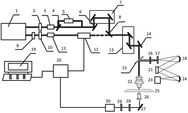
[0020] Such as figure 1 As shown, a molecular vibration spectrum detection and imaging device with different modes includes a laser system 1, and the output direction of the laser system 1 is sequentially provided with a first pulse laser power adjustment unit, an optical path switching device 4, and a pulse laser along the first optical path. A stretching device 5 and a time delay unit 7, the output direction of the laser system 1 is sequentially provided with a second pulse laser power adjustment unit and an acousto-optic modulator 12 along the second optical path, the time delay unit 7 and the second pulse laser power The output direction of the adjustment unit is jointly provided with the first dichroic mirror 8, the laser beam scanning unit 13, the second dichroic mirror 15, the first objective lens 22 and the stage for placing the sample to be measured 25 in sequence. The output end of the second dichroic mirror 15 is sequentially provided with a first converging lens 16...


PUM


Abstract

The invention relates to a different modal molecular vibration spectrum detection and imaging device and a method. The device mainly consists of related apparatuses of functional units such as a laser unit, an optical scanning microscopic unit, a stimulated Raman scattering signal probing and acquiring unit and a spontaneous Raman spectrum detection member and the like. In light of implementation by the method, two different modal molecular vibration scattered signals are quickly acquired in situ on a living cell, an isolated biological tissue and a living small animal: spontaneous Raman spectrum detection under a single laser beam working mode and stimulated Raman scattering microimaging under a double laser beam working mode to achieve quantitative microimaging and qualitative spectral analysis on a target so as to obtain characteristic information of target components, thereby providing important data for optical analysis and deep analysis of a sample.

Description

technical field [0001] The invention relates to a different-mode molecular vibration spectrum detection and imaging device and method. Background technique [0002] Optical microscopy imaging technology plays an important role in the field of biology due to its non-destructive and non-invasive advantages to biological samples. In particular, laser confocal scanning microscopy imaging and two-photon excitation fluorescence imaging technologies that emerged at the end of the nineteenth century, due to their high spatial resolution and three-dimensional optical imaging capabilities, have made optical microscopy widely used in biology, The basic research and clinical application of medicine and related interdisciplinary subjects have been widely used. However, exogenous fluorescent dyes are usually needed for fluorescence microscopy imaging of biological samples, and the addition of exogenous dyes will interfere and affect the characteristics of some small molecules to a certai...

Claims


Application Information

Patent Timeline
Patent Type & Authority Applications(China)
IPC IPC(8): G01N21/65
Inventor 杨洪钦邱彩敏陈建玲彭亦如王瑜华郑莉琴谢树森
Owner FUJIAN NORMAL UNIV
Why Eureka
Try Eureka