Method for preparing cartilage with high mechanical property based on 3D bioprinting

A bioprinting and cartilage technology, applied in the field of 3D bioprinting, can solve the problems of poor mechanical properties and low cell survival rate, and achieve the effect of improving mechanical properties, excellent mechanical properties, and not easy to break

Active Publication Date: 2015-03-11
西安博恩生物科技有限公司
View PDF4 Cites 38 Cited by
  • Summary
  • Abstract
  • Description
  • Claims
  • Application Information

AI Technical Summary

Problems solved by technology

[0003] The purpose of the present invention is to provide a method for preparing cartilage with high mechanical properties based on 3D bioprinting to solve the problems of poor mechanical properties and low cell survival rate in bionic cartilage in the prior art

Method used


Image

  • Method for preparing cartilage with high mechanical property based on 3D bioprinting
  • Method for preparing cartilage with high mechanical property based on 3D bioprinting

Examples


Embodiment 1

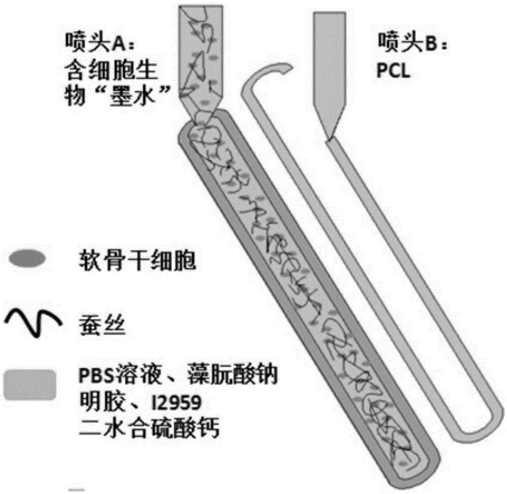
[0022] (1) Preparation of silk fiber: Cut the cocoon into fragments of 0.5 cm in length, put the fragments in a sodium carbonate solution with a molar concentration of 0.02M, stir and boil for 40 minutes, rinse the silk three times for 20 minutes, take out the tweezers and dry overnight. Drop the 10.0M lithium bromide solution on the silk and incubate at 70℃ for 3h. Put the cultured silk into a dialysis box and dialyze it in ultrapure water for 24h. Take out the silk solution in the dialysis box and centrifuge twice at 4℃. Next store.

[0023] (2) Preparation of gelatin solution: add 7.0%wt of phosphate buffer to the phosphate buffer, stir continuously at 45°C until the gelatin is completely dissolved, add 1.5mL to the solution while stirring at a speed of 0.5mL / min A methacrylic anhydride solution with a mass concentration of 94% was reacted at 50℃ for 1h, then diluted with 40mL phosphate buffer at 40℃, and then dialyzed with 12000 dialysis membrane in 40℃ deionized water for 1 ...

Embodiment 2

[0029] (1) Preparation of silk fiber: Cut silkworm cocoons into fragments with a side length of 1.0cm, put the fragments in a sodium carbonate solution with a molar concentration of 0.02M, stir and boil for 50min, rinse the silk three times for 20min, take out the tweezers and dry overnight. Drop the 7.0M lithium bromide solution on the silk and incubate at 50℃ for 5h. Put the cultured silk into a dialysis box and dialyze in ultrapure water for 48h. Take out the silk solution in the dialysis box and centrifuge twice at 4℃. Next store.

[0030] (2) Preparation of gelatin solution: add 10.0%wt of phosphate buffer mass gelatin to the phosphate buffer, stir continuously at 60℃ until the gelatin is completely dissolved, add 0.5mL to the solution while stirring at a speed of 1.0mL / min A methacrylic anhydride solution with a mass concentration of 94% was reacted at 50°C for 1 hour, then diluted with 40mL phosphate buffer at 40°C, and then dialyzed with a 14000 dialysis membrane in 40°C ...

Embodiment 3

[0036] (1) Preparation of silk fiber: Cut the cocoon into pieces with a side length of 0.8cm, put the pieces in a sodium carbonate solution with a molar concentration of 0.02M, stir and boil for 30 minutes, rinse the silk three times for 20 minutes, take out the tweezers and dry overnight. Drop the 12.0M lithium bromide solution on the silk and incubate it at 30℃ for 2h. Put the cultured silk into a dialysis box and dialyze it in ultrapure water for 60h. Take out the silk solution in the dialysis box and centrifuge twice at 4℃. Next store.

[0037] (2) Preparation of gelatin solution: add 12%wt of phosphate buffer to the phosphate buffer, stir continuously at 30°C until the gelatin is completely dissolved, and add 1 mL of weight to the solution while stirring at a speed of 0.1mL / min 94% methacrylic anhydride solution, react at 50℃ for 1h, then dilute with 40mL phosphate buffer at 40℃, then dialyzed with 13000 dialysis membrane in 40℃ deionized water for 1 week, and finally use th...


PUM


Abstract

The invention provides a method for preparing cartilage with a high mechanical property based on 3D bioprinting. The method comprises the following steps: preparing silk fibers, preparing a gelatin solution, preparing ciological ink containing cells, carrying out 3D cartilage printing, performing cell culture and differentiation on a printed tissue to form the cartilage. The method solves the problems of polarization of 2-dimension culture for cells and high body and surface ratio of cells through the 3D bioprinting technology; sodium alginate and calcium ions form ionic bonds, and gelatin and methacrylic anhydride form covalent bonds, so that the 3D bioprinted cartilage is good in mechanical property and is not easily broken; the gelatin has biocompatibility and degradability of normal biological material hydrogel, has bioactivity, and is good for cell growth and differentiation and realization of cell functions.

Description

technical field [0001] The invention belongs to the technical field of 3D bioprinting, and particularly relates to a method for preparing cartilage with high mechanical properties and an environment favorable for cell growth and differentiation based on 3D bioprinting. Background technique [0002] At present, most of the scaffold materials used in bionic cartilage research are gel materials. PVA and PEG have been extensively studied due to their non-toxic, hydrophilic, degradable and cytocompatibility, but these hydrogels have small tensile fracture energy. About 10Jm ‐2 (Cartilage is about 1000Jm ‐2 ), the cracks in the gel have a great influence on its mechanical properties, and the mechanical properties are poor; the synthetic hydrogels on the market have excellent mechanical properties, the tensile fracture energy is large, and the cracks in the gel have little influence on their mechanical properties, but these hydrogels Glue cannot be used for cell culture; none of ...

Claims


Application Information

Patent Timeline
Patent Type & Authority Applications(China)
IPC IPC(8): A61L27/22A61L27/52
Inventor 袁方臧剑锋戴祖明裴梦婷黄琦
Owner 西安博恩生物科技有限公司
Why Eureka
Try Eureka