

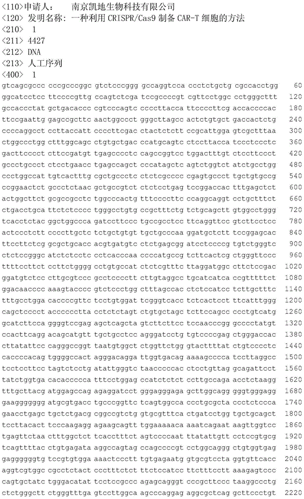
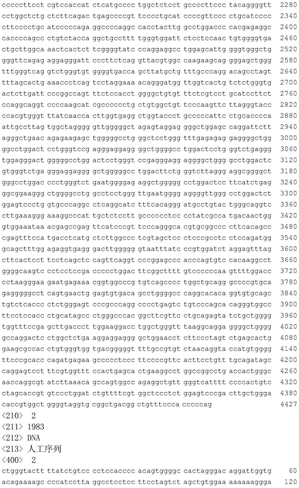
Method for preparing CAR-T cell by CRISPR/Cas9
A cell and DNA sequence technology, applied in the field of biology, to achieve the effect of high editing efficiency, avoiding potential safety hazards and high safety
- Summary
- Abstract
- Description
- Claims
- Application Information
AI Technical Summary
Problems solved by technology
Method used
Image

Examples
Embodiment 1
[0020] Embodiment 1 (preparation embodiment)
[0021] Production of leukemia CAR-T cells using CRISRP / Cas9 system:
[0022] 1) T cell isolation and culture
[0023] a. Separation of fresh peripheral blood mononuclear cells (PBMC, peripheral blood mononuclear cell) by density gradient centrifugation;
[0024] b. Use paramagnetic beads (Dynabeads ClinExVivo CD3 / CD28, Invitrogen, Camarillo, CA, USA) coupled with anti-CD3 and anti-CD28 antibodies to enrich CD3+ cells. The ratio of magnetic beads to cells is 3:1; cells are diluted to TNC (total nucleated cell) at a concentration of 20-30x106 / mL, co-incubate with magnetic beads in a petri dish for 2 hours at room temperature;
[0025] c. Use Magnetic particles concentrator (MPC) (Invitrogen) to enrich CD3+ cells; cells containing CD3+ are resuspended in culture medium (OpTmizer TM CTS TMT-Cell Expansion SFM, Life Technologies), the final concentration was 1×10⁶cells / mL. Incubate for 2 days at 37°C in a 5% CO2 incubator.
[00...
PUM
 Abstract
Description
Claims
Application Information