Cell recognition apparatus and method based on phase contrast image and confocal scattering microspectrum

A phase contrast image and cell identification technology, which is applied in the direction of using optical devices, measuring devices, color/spectral characteristic measurement, etc.

Inactive Publication Date: 2015-11-18
UNIV OF SHANGHAI FOR SCI & TECH
View PDF4 Cites 2 Cited by
  • Summary
  • Abstract
  • Description
  • Claims
  • Application Information

AI Technical Summary

Problems solved by technology

[0004] Medical theory and clinical diagnostic practice and research results show that the gold standard for tumor diagnosis is microscopic observation of pathological sections, but microscopic images only reflect the morphological characteristics of cells. On the other hand, analysis is time-consuming and laborious, and subjective factors such as doctors' clinical experience have a greater impact on the diagnosis
There are some defects in the currently developed detection methods based on optical methods: endoscopic laser confocal microscopy, as a current research hotspot, can only obtain the changes in the three-dimensional morphology of tissue cells, and give qualitative disease analysis, which is different from traditio

Method used


Image

  • Cell recognition apparatus and method based on phase contrast image and confocal scattering microspectrum
  • Cell recognition apparatus and method based on phase contrast image and confocal scattering microspectrum

Examples


Embodiment Construction

[0026] In order to make the technical means, creative features, goals and effects of the present invention easy to understand, the following embodiments will specifically illustrate the cell identification device based on phase contrast images and confocal scattering microspectroscopy of the present invention in conjunction with the accompanying drawings.

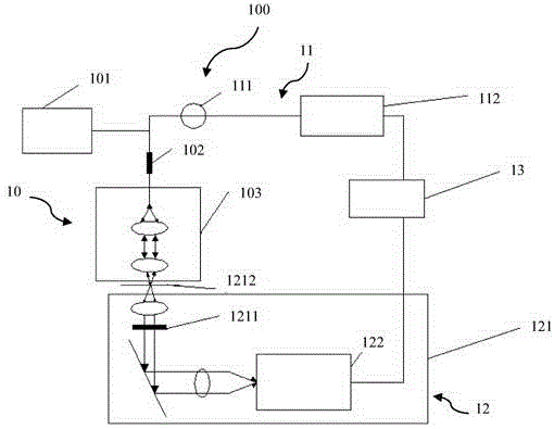
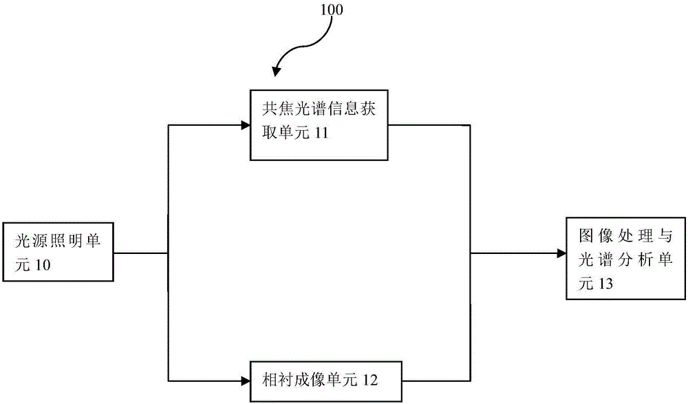
[0027] figure 1 It is a schematic diagram of the composition of the cell identification device based on phase contrast image and confocal scattering microspectroscopy in this embodiment.

[0028] Such as figure 1 As shown, the cell identification device 100 based on phase contrast images and confocal scattering microspectroscopy includes:

[0029] a light source lighting unit 10 for emitting and transmitting light,

[0030] A confocal spectrum information acquisition unit 11, configured to collect another part of the parallel light scattered by the sample to obtain a spectrum containing the cell component information;

...


PUM


Abstract

The invention provides a cell recognition apparatus based on phase contrast image and confocal scattering microspectrum. The cell recognition apparatus is used for detecting early stage cancerous cells in a sample to be detected, and comprises: a light source illumination unit, which is used for emitting and transmitting light; a phase contrast imaging unit, which comprises a phase contrast microscope and a camera; a confocal spectrum information obtaining unit, which comprises a spectrometer for collecting light which is scattered from the sample to be detected, wherein, the light that irradiates the sample is from the other part of light that penetrates from a coupling lens set and the light scattered from the sample is received and transmitted by the coupling lens set again, and obtaining a spectrum containing the component information of the cells; an image processing and spectral analysis unit, which is used for receiving the phase contrast image and the spectrum, and calculating, analyzing and obtaining a qualitative and a quantitative matching result between cells in the sample to be detected and the normal cells; and a control unit for controlling the light source illumination unit, the phase contrast imaging unit, the confocal spectrum information obtaining unit and the image processing and spectral analysis unit to work normally.

Description

technical field [0001] The invention relates to a device and method for detecting cells, in particular to a cell identification device and method capable of simultaneously obtaining cell morphology and principal components. Background technique [0002] Early cancer detection is a major topic in the medical field. Traditional detection techniques can only detect tumors when they reach a certain size, which often delays the optimal treatment time. If it can be detected when a small amount of cancer changes in the early stage of cells, it will greatly help cancer patients and doctors to detect, intervene and treat early, and improve the survival time and quality of life of cancer patients. [0003] In the process of biological tissue canceration, the shape and structure of cancerous cells are very different from normal cells. These differences are reflected in: the volume of cancer cells is larger than that of normal cells, the nucleus is also significantly enlarged and irreg...

Claims


Application Information

Patent Timeline
IPC IPC(8): G01N21/84G01N21/25G01B11/24
Inventor 王成杨静张通郭旭东
Owner UNIV OF SHANGHAI FOR SCI & TECH
Why Eureka
Try Eureka