Multimodal particles, methods and uses thereof
A technology of particles, uses, applied in the field of multimodal particles, their uses and uses
- Summary
- Abstract
- Description
- Claims
- Application Information
AI Technical Summary
Problems solved by technology
Method used
Image
Examples
example 1
[0168] Example 1: Synthesis of SE(R)RS Particles
[0169] by adding 20 mM HAuCl at 4 °C 4Gold nanostar substrates were synthesized by rapid addition to 40 mM ascorbic acid. Ascorbate-stabilized gold nanostars (about 75 nm, 1 nM) thus synthesized were collected by centrifugation (3,500 xg, 15 min) and dialyzed overnight. Dialyzed gold nanostars were coated with dye-intercalated silica by the typical Stobel method. Briefly, dialyzed gold nanostars were added to ethanol (added with resonance Raman dye, TEOS and ammonia) and allowed to react for 1 hour. Particles were isolated by centrifugation (3,500 xg, 15 min) and washed with ethanol. To achieve PEGylation, the silica surface was modified with mercapto groups by heating the silica-coated nanostars in ethanol containing 1% (v / v) MPTMS at 72°C for 1 hour. The nanostars were washed with ethanol to remove MPTMS and redispersed in 1% (w / v) methoxy-terminated (m)PEG 2000 - maleimide in 10 mM MES buffer (pH 7.1). maleimide-mPEG ...
example 2
[0171] Example 2: Characterization
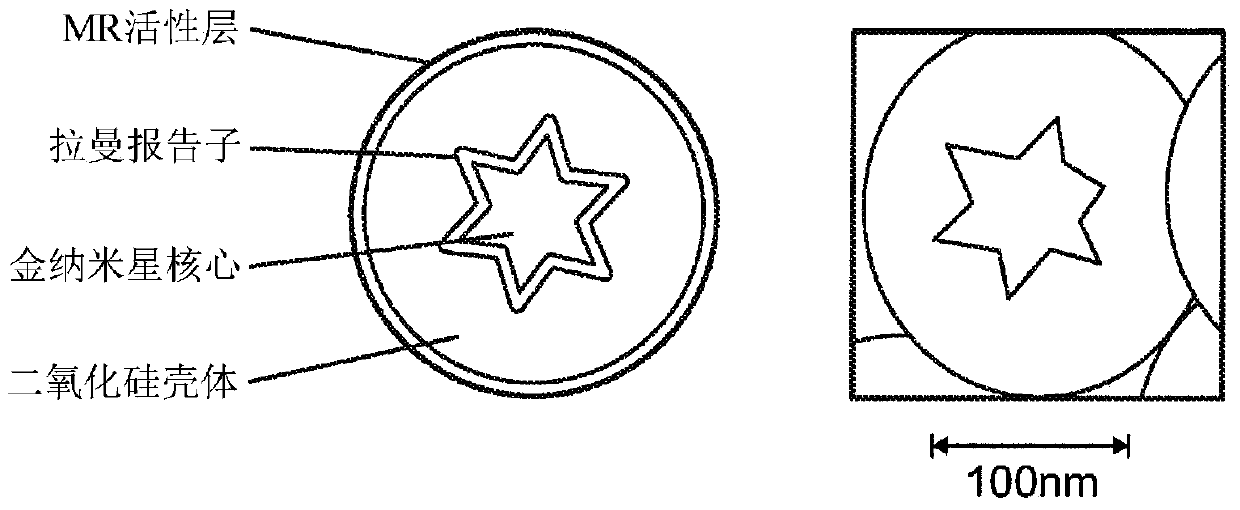
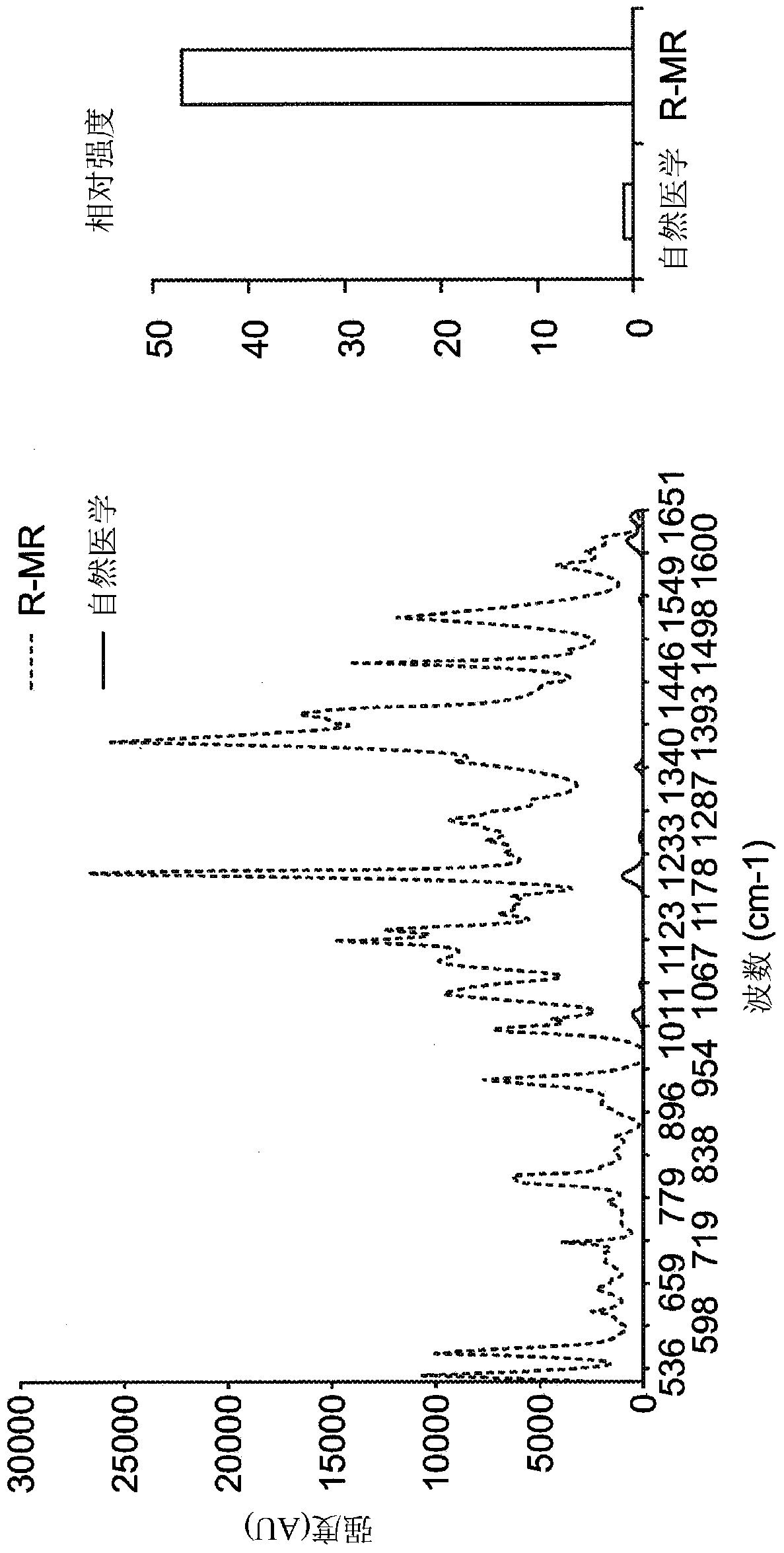
[0172] Ultra-high sensitivity: such as figure 2 As shown in , the SE(R)RS particles synthesized in Example 1 were characterized by transmission electron microscopy (TEM; JEOL 1200EX (JEOL 1200EX); USA), size distribution, and by nanoparticle tracer analysis (NTA; Nanosight (Nanosight, UK) was used to determine the concentration. Equipped with a 300mW 785nm (near-IR) diode laser and a 1-inch charge-coupled device detector (spectral resolution of 1.07cm -1 Raman activity of equimolar amounts of particles was measured on a Renishaw InVIA Raman microscope (Renishaw InVIA Raman microscope). Raman spectra were analyzed with WiRE 3.4 software (Renishaw, UK).
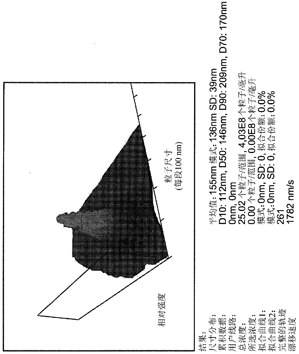
[0173] Nanoparticle Tracer Analysis (NTA): eg image 3 The size distribution of 1 pM particles in water was determined by NTA as shown in .
example 3
[0174] Example 3: Animal Testing
[0175] refer to Figure 4-10 , injected tumor-bearing mice (dedifferentiated liposarcoma model, PyMT-MMTV (fvb) transgenic breast cancer model, Hi-MYC transgenic prostate cancer model, RCAS) with 150 μL 2.5nM SE(R)RS particles synthesized in Example 1 / TV-a transgenic glioma model). Animals were sacrificed 18 hours later or later and scanned for Raman activity on the system described above. Tumors, organs and lymph nodes were harvested and additionally imaged ex vivo and then embedded in wax. Embedded tissues were processed for histology (H&E staining, tumor marker staining, macrophage staining).
[0176] In Vivo-Ex Vivo Multimodal MRI-Raman Histology Correlation: As confirmed by the experimental results described below, SE(R)RS particles were able to reliably and with microscopic precision delineate three different xenografted mouse sarcomas Presence of tumors in models (n=5 per model). The cells implanted in these mouse models were der...
PUM
| Property | Measurement | Unit |
|---|---|---|
| size | aaaaa | aaaaa |
| thickness | aaaaa | aaaaa |
Abstract
Description
Claims
Application Information
 - R&D
- Intellectual Property
- Life Sciences
- Materials
- Tech Scout
- Unparalleled Data Quality
- Higher Quality Content
- 60% Fewer Hallucinations
Browse by: Latest US Patents, China's latest patents, Technical Efficacy Thesaurus, Application Domain, Technology Topic, Popular Technical Reports.
© 2025 PatSnap. All rights reserved.Legal|Privacy policy|Modern Slavery Act Transparency Statement|Sitemap|About US| Contact US: help@patsnap.com


