
Nuclear magnetic resonance detection method for biological tissue

A biological tissue and nuclear magnetic resonance technology, applied in the field of nuclear magnetic resonance spectroscopy detection, can solve the problems of internal structure damage of biological tissue, limitations of direct detection, complex sample pretreatment, etc., achieve rapid sampling and overcome the effect of magnetic field inhomogeneity

Active Publication Date: 2015-12-16
XIAMEN UNIV
View PDF7 Cites 17 Cited by
  • Summary
  • Abstract
  • Description
  • Claims
  • Application Information

AI Technical Summary

Problems solved by technology

However, the magic angle rotation experiment requires special probe detection equipment, and high-speed rotation will often cause damage to the internal structure of biological tissues, which cannot guarantee the integrity of the tissue during the experiment
[0004] It can be seen that the conventional two-dimensional J-decomposition spectroscopy method is difficult to be directly used in the detection and analysis of biological tissue metabolism, although the combination of tissue extraction and magic-angle rotation technology can well eliminate the influence of non-uniform magnetic susceptibility inside the tissue to obtain the required analysis results. However, these techniques require complex sample pretreatment and special hardware equipment, and have great limitations in the direct detection of intact biological tissues

Method used


Image

  • Nuclear magnetic resonance detection method for biological tissue
  • Nuclear magnetic resonance detection method for biological tissue
  • Nuclear magnetic resonance detection method for biological tissue

Examples


specific Embodiment

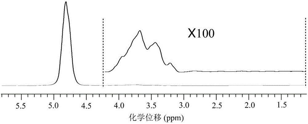
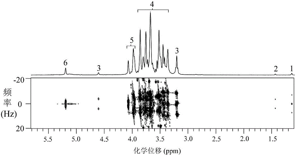
[0041] The method proposed in the present invention is used to scan a biological tissue as an example, and this specific example is used to verify the feasibility of the present invention in the application of biological tissue. The biological tissue sample used in the experiment was grape pulp tissue, and the experimental test was carried out under a Varian 500MHz NMR spectrometer (Varian, Palo Alto, CA). The whole experimental process did not carry out any sample pretreatment on the grape pulp tissue, and no artificial homogenization was carried out. On-site operation, no changes to any instrument hardware facilities. According to the operation process of the method proposed in the present invention, first obtain a one-dimensional spectrum with conventional simple one-dimensional pulse sequence sampling, the sampling time is 4s, the result is as follows figure 2 As shown, the spectral line width can be obtained from this one-dimensional spectrum with a linewidth of 90 Hz, a...


PUM


Abstract

The invention discloses a nuclear magnetic resonance detection method for biological tissue, and relates to a nuclear magnetic resonance spectroscopy detection method. A biological tissue sample to be detected is put into a nuclear magnetic test tube, and then the test tube containing the sample is put into a detection magnetic body of a nuclear magnetic spectrometer; a one-dimensional spectrum is sampled through a conventional one-dimensional pulse sequence, the width of the spectral line is obtained to provide a basis for setting a spectral width parameter, and meanwhile a line width value reflects the nonuniformity situation of a magnetic filed of the biological tissue sample; a pulse sequence which is well compiled in advance is introduced onto a nuclear magnetic resonance spectrometer; an intermolecular zero-quantum coherent signal selection module, a folding-correction module, a solvent suppression module and a J modulating signal sampling module of the pulse sequence are started, and experiment parameters of all the modules are set; after setting of the experiment parameters is completed, the artificial shimming operation process is skipped, and data sampling is directly executed; after data sampling is completely completed, related data post-processing is conducted, and a high-resolution two-dimensional J resolution spectrum which is free of the influence of a nonuniform magnetic field is obtained.

Description

technical field [0001] The invention relates to a nuclear magnetic resonance spectroscopy detection method, in particular to a nuclear magnetic resonance detection method for biological tissue. Background technique [0002] Nuclear Magnetic Resonance (NMR, Nuclear Magnetic Resonance) spectroscopy can provide molecular-level information such as chemical shifts and J-couplings, and has become a powerful tool for molecular structure analysis and component analysis, and is widely used in biological detection and metabolite analysis. Generally, one-dimensional NMR spectroscopy has a short sampling time and can meet the analysis requirements of samples with simple chemical composition and molecular structure, so it is widely used in simple sample systems. However, biological samples often contain a large number of metabolite components and complex molecular structures, which can generate a large number of NMR signals, making the obtained one-dimensional NMR spectrum peaks crowded ...

Claims


Application Information

Patent Timeline
IPC IPC(8): G01N24/08
Inventor 黄玉清汪凯宇陈忠
Owner XIAMEN UNIV
Learn More