Ovarian cancer tissue 3D cultivation method and application of 3D cultivated cancer tissue to efficacy evaluation

A culture method and cancer tissue technology, applied in the field of biology, can solve problems such as long evaluation time, impact of species differences on evaluation results, and inability to accurately reflect drug efficacy, so as to make up for weak links, promote research and development, and achieve precise treatment. The effect of the process

Inactive Publication Date: 2016-01-06
JOINN LAB (SUZHOU) INC
View PDF2 Cites 12 Cited by
  • Summary
  • Abstract
  • Description
  • Claims
  • Application Information

AI Technical Summary

Problems solved by technology

[0007] The main technical problem to be solved by the present invention is to provide a 3D culture method of ovarian cancer tissue and its application in drug efficacy evaluation, which is expected to solve the imp...

Method used


Image

  • Ovarian cancer tissue 3D cultivation method and application of 3D cultivated cancer tissue to efficacy evaluation
  • Ovarian cancer tissue 3D cultivation method and application of 3D cultivated cancer tissue to efficacy evaluation
  • Ovarian cancer tissue 3D cultivation method and application of 3D cultivated cancer tissue to efficacy evaluation

Examples


Embodiment 1

[0033] A method for 3D culture of ovarian cancer tissue is provided, comprising the steps of:

[0034] (1) Preparation of tumor tissue blocks

[0035] Take tumor tissue and cut it into 0.5-1mm with ophthalmic surgical instruments 3 The tissue pieces were washed with phosphate buffered saline (1×PBS), then soaked in DMEM medium and kept on ice for later use;

[0036] (2) Tumor tissue block culture

[0037] Take a 6-well plate, 24-well plate or 96-well plate, add 10-50 μL of diluted Matrigel (1:1-1:5 with DMEM) dropwise along the bottom edge, room temperature or CO 2 Place in the incubator for 10-30min, carefully move the tissue block onto the solidified Matrigel, and then drop 10-50 μL of diluted Matrigel glue (the dilution ratio with DMEM is 1:1-1:5) on the tissue block, Set CO 2 Stand in the incubator for 20-30min.

[0038]Take out the orifice plate, carefully add 0.4mL of culture solution to 24-well plate, 0.1mL to 96-well plate or 2.5mL to 6-well plate for cultivation,...

Embodiment 2

[0044] Feasibility analysis of 3D cultured tumor tissue for efficacy evaluation of antitumor drugs

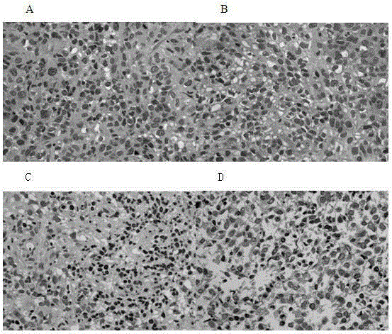
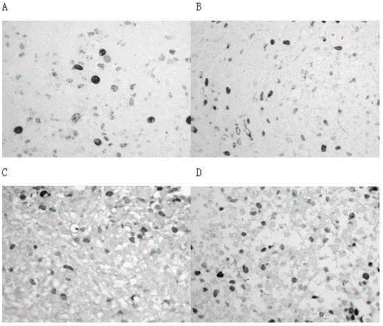
[0045] When ovarian cancer tissue is cultured, different concentrations and different types of chemotherapeutic drugs are added to the culture medium, and at different times after culture, the tissue is taken out, frozen sections are made or fixed in the above-mentioned fixative solution, and stained by HE, Ki67 immunohistochemical staining or Corresponding to other staining, observe the tissue structure and cell morphology under the microscope, especially the changes in the shape of the nucleus and the depth of staining, randomly count the number of cells with distorted nuclei in the total number of cells (percentage), and compare with those without antitumor drugs The difference in the nuclear aberration rate of the specimens is used to evaluate the efficacy of the drug. For details, see figure 2 and Figure 4 .

[0046] figure 2 Changes in the tissue structure and nucle...


PUM


Abstract

The invention discloses an ovarian cancer tissue 3D cultivation method. The method includes the steps that matrigel is dripped into a pore plate, and standing is conducted at an indoor temperature or in a CO2 cultivation box so that the gel can be solidified; a tumor tissue block submerged in a cultivation solution is moved onto the solidified matrigel, then matrigel is dripped onto the tumor tissue block and the gel is solidified in the same way; a cultivation solution is added for cultivation for 1-3 days; the tumor tissue block is taken out and fixed. Chemotherapeutics can be added during cultivation, the tissue block is fixed after cultivation, the cell shape is observed after dyeing, and the nucleus malformation rate is calculated. By means of the established cultivation method and detection indexes, the pharmacological effects and the function characteristics of anti-tumor drugs can be evaluated more quickly and accurately, the technical defects of cell 2D cultivation and animal experimental evaluation can be overcome, and it is hopeful that the research and development of innovative anti-cancer drugs in China and the precision therapeutic process can be promoted.

Description

technical field [0001] The invention relates to the field of biology, in particular to a 3D culture method for ovarian cancer tissue and its application in drug efficacy evaluation. Background technique [0002] Tumor is one of the major diseases that seriously endanger people's health. The pathogenesis of tumors is diverse and complex. However, the current conventional chemotherapeutic drugs are almost all cytotoxic drugs targeting cells with strong proliferative ability. toxic side effect. Therefore, many drug research and development institutions are committed to the development of highly efficient, low-toxic and highly specific targeted anti-tumor drugs, and the traditional single anti-tumor drug screening method is no longer enough to meet the needs of the development of anti-tumor drug pharmacodynamics. [0003] With the deepening understanding of tumor pathogenesis and the development of tumor-targeted drugs, precision medicine has gradually become a trend in the res...

Claims


Application Information

Patent Timeline
IPC IPC(8): C12N5/09C12Q1/02
Inventor 李月娟易月娥顾阳陆益彬陈海宁左从林孙云霞冯宇霞
Owner JOINN LAB (SUZHOU) INC
Why Eureka
Try Eureka