Preparation method for CIK cells with high cytotoxic activity
A cytotoxic and cellular technology, applied in the field of cellular immunity, can solve the problems of impaired immune function, weakened immune cell function, and low ability to kill tumors, and achieve the effects of high proliferation, high purity, and high cytotoxic activity.
- Summary
- Abstract
- Description
- Claims
- Application Information
AI Technical Summary
Problems solved by technology
Method used
Image
Examples
Embodiment 1
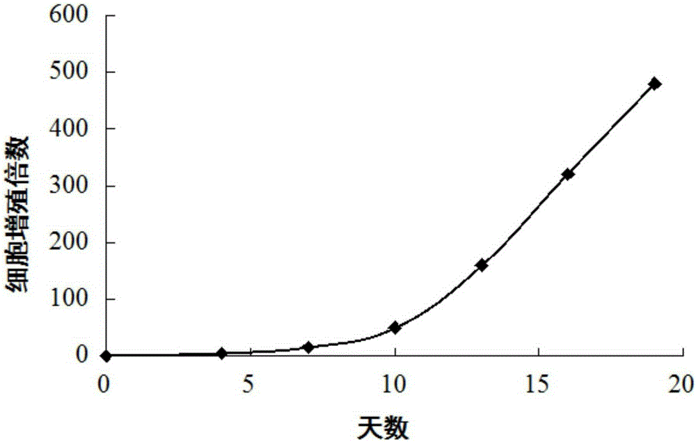
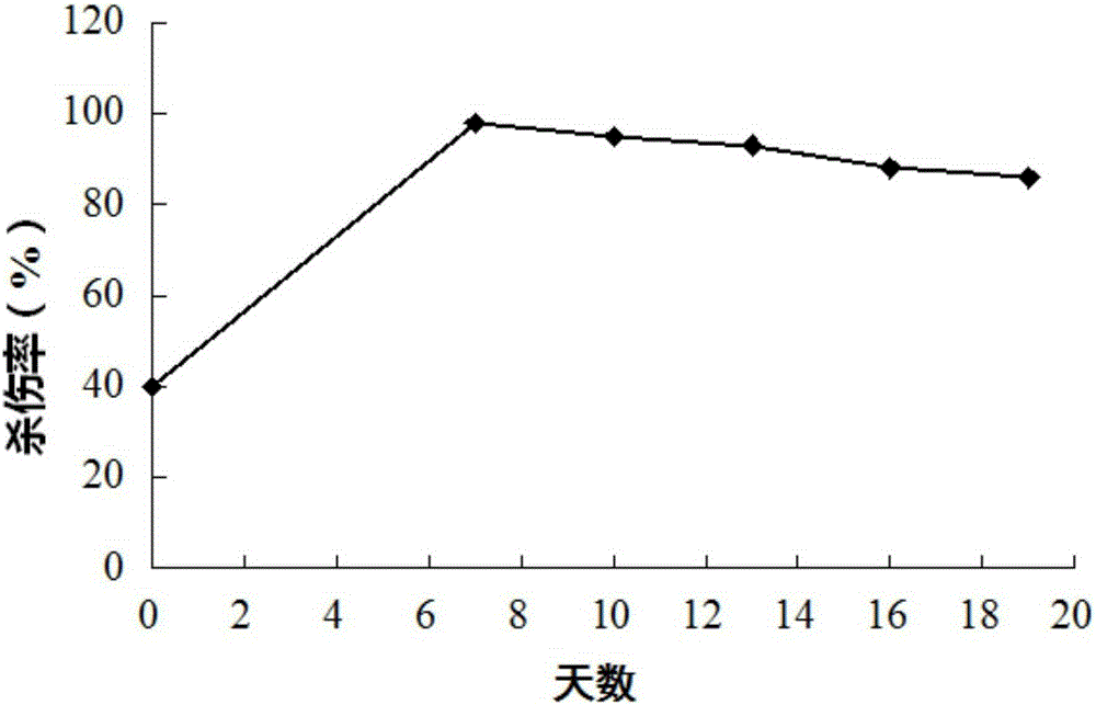
[0020] Example 1: Induced preparation and detection of CIK cells
[0021] 1. Induction and preparation of CIK cells
[0022] (1) Take 30 mL of peripheral blood from the patient and add an equal volume of RPMI1640 culture medium (Invitrogen Company), and mix well.
[0023] (2) Add 60 mL of the doubly diluted peripheral blood obtained in step (1) to the interface of 30 mL of lymphocyte separation medium (Shanghai Huajing Biological High-tech Co., Ltd.) (specific gravity 1.077) (do not destroy the interface), and use 2000 rpm, centrifuge for 20 minutes, take the middle layer - the buffy coat layer rich in lymphocytes, and add 20 mL of complete medium. A small amount of cells were taken for cell counting and trypan blue live cell count staining, and the results showed that the live cells reached 99%.
[0024] (3) Centrifuge the buffy coat layer containing complete medium obtained in step (2) at 1000 rpm for 10 minutes, discard the supernatant, and precipitate mononuclear cells. ...
Embodiment 2
[0050] Example 2: Induced preparation of CIK cells
[0051] (1) Take 30 mL of peripheral blood from the patient and add an equal volume of RPMI1640 culture medium (Invitrogen Company), and mix well.
[0052] (2) Add 60 mL of the doubly diluted peripheral blood obtained in step (1) to the interface of 30 mL of lymphocyte separation medium (Shanghai Huajing Bio-Technology Co., Ltd.) (specific gravity 1.077) (do not destroy the interface), and use 1500 rpm, centrifuge for 20 minutes, take the middle layer - the buffy coat layer rich in lymphocytes, and add 20 mL of complete medium. A small amount of cells were taken for cell counting and trypan blue live cell count staining, and the results showed that the live cells reached 99%.
[0053] (3) Centrifuge the buffy coat layer containing complete medium obtained in step (2) at 1500 rpm for 7 minutes, discard the supernatant, and precipitate mononuclear cells.
[0054] (4) Add complete medium, count the cells, adjust the mononuclea...
Embodiment 3
[0061] Example 3: Induced preparation and detection of CIK cells
[0062] (1) Take 30 mL of peripheral blood from the patient and add an equal volume of RPMI1640 culture medium (Invitrogen Company), and mix well.
[0063] (2) Add 60 mL of the doubly diluted peripheral blood obtained in step (1) to the interface of 30 mL of lymphocyte separation medium (Shanghai Huajing Bio-Technology Co., Ltd.) (specific gravity 1.077) (do not destroy the interface), and use 2500 rpm, centrifuge for 15 minutes, take the middle layer - the buffy coat layer rich in lymphocytes, and add 20 mL of complete medium. A small amount of cells were taken for cell counting and trypan blue live cell count staining, and the results showed that the live cells reached 99%.
[0064] (3) Centrifuge the buffy coat layer containing complete medium obtained in step (2) at 1500 rpm for 7 minutes, discard the supernatant, and precipitate mononuclear cells.
[0065] (4) Add complete medium, count the cells, adjust ...
PUM
 Abstract
Description
Claims
Application Information
 - R&D
- Intellectual Property
- Life Sciences
- Materials
- Tech Scout
- Unparalleled Data Quality
- Higher Quality Content
- 60% Fewer Hallucinations
Browse by: Latest US Patents, China's latest patents, Technical Efficacy Thesaurus, Application Domain, Technology Topic, Popular Technical Reports.
© 2025 PatSnap. All rights reserved.Legal|Privacy policy|Modern Slavery Act Transparency Statement|Sitemap|About US| Contact US: help@patsnap.com


