Fluorescence light sheet microscopy imaging system and method

A technology of microscopic imaging and fluorescent light sheet, which is applied in the field of microscope systems and can solve the problems of unsuitable wavefront distortion sensing and correction methods.

Active Publication Date: 2018-02-13
PEKING UNIV
View PDF8 Cites 0 Cited by
  • Summary
  • Abstract
  • Description
  • Claims
  • Application Information

AI Technical Summary

Problems solved by technology

However, unlike the two-photon microscope in the point-by-point scanning mode, the light-sheet microscope in the layer-by-layer scanning mode is not suitable for the above-mentioned wavefront distortion sensing and correction method

Method used


Image

  • Fluorescence light sheet microscopy imaging system and method
  • Fluorescence light sheet microscopy imaging system and method
  • Fluorescence light sheet microscopy imaging system and method

Examples


Embodiment Construction

[0029] In the drawings, the same or similar reference numerals are used to denote the same or similar elements or elements having the same or similar functions. Embodiments of the present invention will be described in detail below in conjunction with the accompanying drawings.

[0030] In the description of the present invention, the terms "central", "longitudinal", "transverse", "front", "rear", "left", "right", "vertical", "horizontal", "top", " The orientation or positional relationship indicated by "bottom", "inner", "outer", etc. is based on the orientation or positional relationship shown in the drawings, and is only for the convenience of describing the present invention and simplifying the description, rather than indicating or implying the referred device or element Must have a specific orientation, be constructed and operate in a specific orientation, and therefore should not be construed as limiting the scope of the invention.

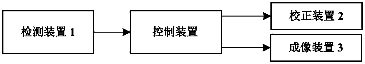
[0031] Such as figure 1 and Figu...


PUM

PropertyMeasurementUnit
thicknessaaaaaaaaaa

Abstract

The invention discloses a fluorescent light sheet microscopic imaging system and method. The fluorescent light sheet microscopic imaging system includes a detection device, a correction device, an imaging device and a control device, wherein: the detection device is used to detect the wavefront of each preset isohalo region of the tissue plane imaging field of view inside the living sample aberration distortion, and send it to the control device, the control device is used to send correction instructions to the correction device according to the received wavefront aberration distortion, and the correction device is used to send correction instructions to the correction device according to the received The correction instruction is used to perform at least one wavefront correction on each of the isohales at the same time, and the imaging device is used to image the tissue plane after wavefront correction. The invention helps to obtain a deep biological tissue plane with a large field of view and high temporal and spatial resolution.

Description

technical field [0001] The invention relates to a microscope system, in particular to a fluorescent light sheet microscopic imaging system and method. Background technique [0002] In 1993, Voie A.H. et al. invented the light sheet microscope (Orthogonalplane Fluorescence Optical Sectioning, OPFOS), which uses a sheet of light to illuminate transparent biological tissues laterally, thereby realizing microscopic imaging of biological tissues at different depths. However, due to the low imaging resolution at that time and the inability to make live cell measurements, it was not used in the ensuing decade. Until 2004, Huisken J. and Stelzer E.H. et al. used a cylindrical lens to focus the incident excitation light on the sample through the incident objective lens to form a static Gaussian light sheet, and then observed it through a high numerical aperture fluorescence objective lens, establishing the first generation of static fluorescence Light Sheet Microscopy (Selective Pla...

Claims


Application Information

Patent Timeline
Patent Type & Authority Patents(China)
IPC IPC(8): G01N21/64
Inventor 毛珩陶乐天姜明陈良怡李海文安捷宗伟健
Owner PEKING UNIV
Why Eureka
Try Eureka