Retinal cell microscopic image segmentation and counting method

A technology of retinal cells and microscopic images, applied in the field of image processing, can solve the problems of difficult selection and matching of pit points, differences in segmentation results, and failure to use grayscale information, etc., achieving good accuracy, small amount of calculation, and simple operation Effect

Inactive Publication Date: 2016-05-25
SUZHOU UNIV
View PDF4 Cites 11 Cited by
  • Summary
  • Abstract
  • Description
  • Claims
  • Application Information

AI Technical Summary

Problems solved by technology

[0003] The current cell image automatic segmentation algorithm has the following defects: (1) Most of the algorithms look for concave points with higher curvature on the contour of cohesive cells as the starting point of segmentation, and there are often multiple concave points on the cell contour, and the number of concave points Difficult to choose and match
(2) Most of the existing s...

Method used


Image

  • Retinal cell microscopic image segmentation and counting method
  • Retinal cell microscopic image segmentation and counting method
  • Retinal cell microscopic image segmentation and counting method

Examples


Embodiment Construction

[0024] The present invention will be further described below in conjunction with the accompanying drawings. The following examples are only used to illustrate the technical solutions of the present invention more clearly, and cannot be used to limit the protection scope of the present invention.

[0025] The specific steps of this method are as follows:

[0026] (1) Image preprocessing:

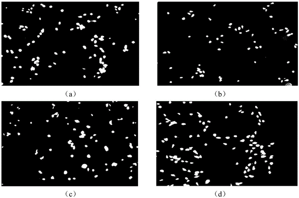
[0027] Since retinal cells are co-cultured with polymer materials, which will lead to irregular cell staining, the acquired fluorescence microscopic images have different degrees of noise. The images are preprocessed by threshold filtering and digital morphological transformation. The filtered image is eroded and expanded using a 3*3 template, which can effectively filter out noise points in the image. Moreover, this method is simple, has low computational cost and high processing efficiency, and can achieve good image denoising and enhancement effects.

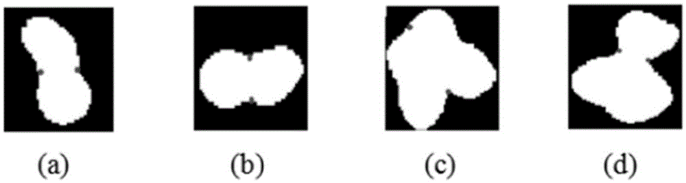
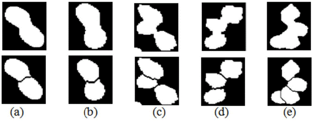
[0028] (2) Shape classification: ...


PUM


Abstract

The invention discloses a retinal cell microscopic image segmentation and counting method. The method is characterized by comprising the following steps: firstly, preprocessing images; secondly, performing shape classification on cell communication regions by using an AdaBoost classifier, and judging whether the segmentation needs to be carried out or not; thirdly, performing bottleneck point detection on cells and connecting segmentation points by using an accelerative Dijkstra algorithm to finish the segmentation; and finally, putting new cell communication regions obtained by segmentation into the AdaBoost classifier again to perform shape classification, and judging whether the segmentation needs to continue to be carried out or not. The method has the beneficial effects that by utilizing the advantages of small calculation amount, simple calculation, high running efficiency and the like in threshold filtering and digital morphology operations, and in combination with cell shape classification and edge profile bottleneck point detection methods and the accelerative Dijkstra algorithm, the overlapped adherent cells are segmented; an obtained result has relatively high accuracy; and the method has relatively high running efficiency.

Description

technical field [0001] The invention relates to a retinal cell microscopic image segmentation and counting method, which belongs to the technical field of image processing. Background technique [0002] The retina is an extension of the neural tissue in the brain and has a complex multi-layered organizational structure. The damage of retinal tissue caused by retinopathy is often difficult to repair by drugs, so biological tissue repair engineering has become a powerful tool for repairing retinal damage. One of the methods is to design a biocompatible material that will carry The material of healthy retinal cells is injected into the damaged part of the retina, which promotes the growth and proliferation of cells in the damaged part, and then repairs the damaged part. This requires this material to be compatible with the human body and harmless to the human body, so the cytotoxicity of this material needs to be accurately assessed. The cytotoxicity of the material was evalu...

Claims


Application Information

Patent Timeline
IPC IPC(8): G06T7/00
CPCG06T2207/10056G06T2207/30041G06T2207/30242
Inventor 陈新建费健峻朱伟芳石霏向德辉林潇杨磊
Owner SUZHOU UNIV
Why Eureka
Try Eureka