Method for establishing in-vitro blood-brain barrier model based on micro-fluidic chip

A microfluidic chip and chip technology, applied in biochemical equipment and methods, artificial cell constructs, tissue cell/virus culture devices, etc., can solve the problems that new microfluidic chips have not yet appeared

Inactive Publication Date: 2016-06-01
DALIAN INST OF CHEM PHYSICS CHINESE ACAD OF SCI
View PDF4 Cites 8 Cited by
  • Summary
  • Abstract
  • Description
  • Claims
  • Application Information

AI Technical Summary

Problems solved by technology

At present, the use of microfluidic chips to construct in vitro blood-brain barrier models is in its infancy, but new microfluidic chips that are close to the in vivo anatomical and physiological microenvironments have not yet appeared.

Method used


Image

  • Method for establishing in-vitro blood-brain barrier model based on micro-fluidic chip
  • Method for establishing in-vitro blood-brain barrier model based on micro-fluidic chip
  • Method for establishing in-vitro blood-brain barrier model based on micro-fluidic chip

Examples


Embodiment 1

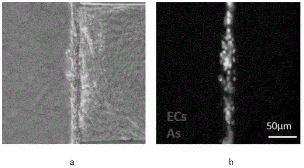
[0035] The primary extracted SD rat brain microvascular endothelial cells (ECs) and brain astrocytes (As) were used to form a blood-brain barrier model at the two-dimensional and three-dimensional microenvironment interface of the microfluidic chip.

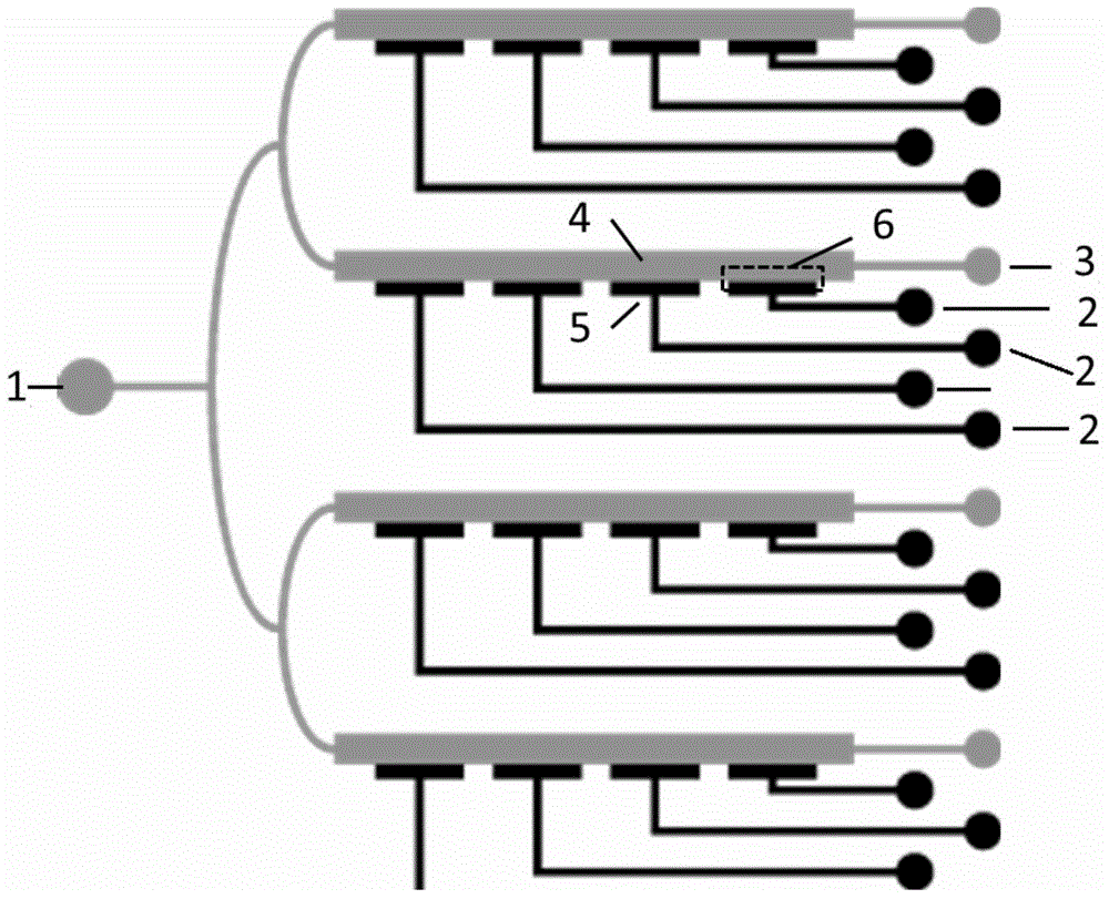
[0036] Apply self-designed and fabricated microfluidic chip, the structure is as follows figure 1 shown. The prepared collagen solution with a concentration of 6mg / mL was poured into the three-dimensional channel from the collagen inlet by a perfusion pump in a continuous perfusion manner, and the culture dish with the fixed chip was placed in a 37°C culture environment to incubate the gel to form a three-dimensional structure. Then the SD rat brain microvascular endothelial cells (ECs) and brain astrocytes (As) that were extracted and cultivated to contact inhibition were respectively digested into single cell suspensions, and mixed according to the ratio of ECs:As=1:1 , use a perfusion pump to inject the mixed cell suspension ...

Embodiment 2

[0038] The resistance value of the blood-brain barrier model structure was measured by an electric resistance measuring instrument, measured once every 2 hours, and the results were as follows: Figure 4 shown. Depend on Figure 4 It shows that with the prolongation of the culture time, the function of the blood-brain barrier model is gradually improved, and the transmembrane resistance is gradually enhanced.

Embodiment 3

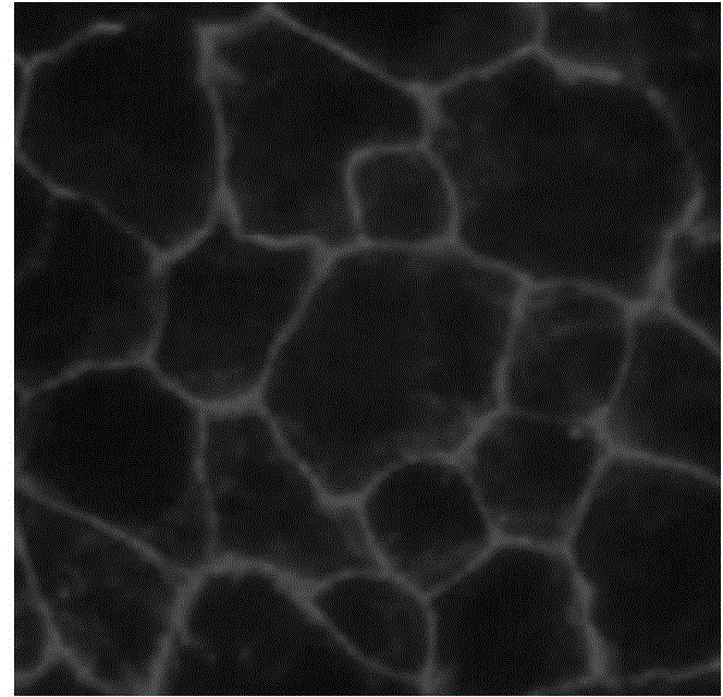
[0040]After establishing the blood-brain barrier model using the method in Example 1, use the perfusion pump to continuously perfuse the CellTracker solution emitting green fluorescence into the two-dimensional channel from the cell inlet, with a concentration of 10 μmol / L and a molecular weight of 557.47 Da, and continuously measure the fluorescence in the three-dimensional channel Intensity, used to characterize the barrier and penetration functions of the blood-brain barrier model to small molecules, the results are as follows Figure 5 shown. Figure 5 It shows that the blood-brain barrier model has a high barrier effect on small molecule fluorescent probes, and the permeability is low.


PUM


Abstract

The invention provides a method for establishing an in-vitro blood-brain barrier model based on a micro-fluidic chip. The micro-fluidic chip mainly comprises a cell inlet, collagen inlets and waste liquid outlets, wherein the cell inlet is communicated with the waste liquid outlets via two-dimensional channels, the collagen inlets are communicated with three-dimensional channels, and two-dimensional and three-dimensional microenvironment interfaces are formed at joints of the two-dimensional channels and the three-dimensional channels. The blood-brain barrier model is established on the base of the micro-fluidic chip. The method comprises the following steps: injection of collagen into the chip; and injection and culture of cells on the chip. Compared with conventional non-contact cell co-culture models or two-dimensional cell culture models, the invention has the following advantages: the method overcomes the problems of secondary inoculation and great time consumption of cell co-culture, solves problems in coexistence of two-dimensional and three-dimensional microenvironments needed by the physiological environment of blood-brain barriers, enables a microenvironment to be closer to the actual physiological microenvironment in the human body and can substantially reduce actual consumption.

Description

technical field [0001] The invention relates to a method using tissue engineering, in particular to a method for establishing an in vitro blood-brain barrier model based on a microfluidic chip. Background technique [0002] The blood-brain barrier is an important structural and functional barrier in mammals, which mainly regulates the entry and exit of molecules into the brain and maintains the homeostasis of the brain microenvironment. Clinical data show that the occurrence and development of various brain diseases are related to the disorder and loss of blood-brain barrier function, and due to the selective penetration of molecules by the blood-brain barrier, many clinical drugs cannot pass the blood-brain barrier smoothly for treatment Therefore, some brain diseases cannot be effectively controlled and treated. Therefore, the establishment of an in vitro blood-brain barrier model with high stability, high reproducibility, and physiological functions close to the body is ...

Claims


Application Information

Patent Timeline
IPC IPC(8): C12M3/00C12N5/071C12N5/079
Inventor 秦建华许慧李中玉
Owner DALIAN INST OF CHEM PHYSICS CHINESE ACAD OF SCI
Why Eureka
Try Eureka