Deep tissue X-ray excitation multispectral tomography system and method

A deep tissue and tomographic imaging technology, which is used in computer tomography scanners, instruments for radiological diagnosis, diagnosis, etc. question

Active Publication Date: 2016-06-08
FOURTH MILITARY MEDICAL UNIVERSITY
View PDF7 Cites 16 Cited by
  • Summary
  • Abstract
  • Description
  • Claims
  • Application Information

AI Technical Summary

Problems solved by technology

[0005] The purpose of the present invention is to provide a system and method for deep tissue X-ray excitation multi-spectral tomography, aiming to solve the problem that most of the emission spectrum of X-ray exc

Method used


Image

  • Deep tissue X-ray excitation multispectral tomography system and method
  • Deep tissue X-ray excitation multispectral tomography system and method

Examples


Embodiment Construction

[0037] In order to make the object, technical solution and advantages of the present invention more clear, the present invention will be further described in detail below in conjunction with the examples. It should be understood that the specific embodiments described here are only used to explain the present invention, not to limit the present invention.

[0038] The application principle of the present invention will be further described below in conjunction with the accompanying drawings.

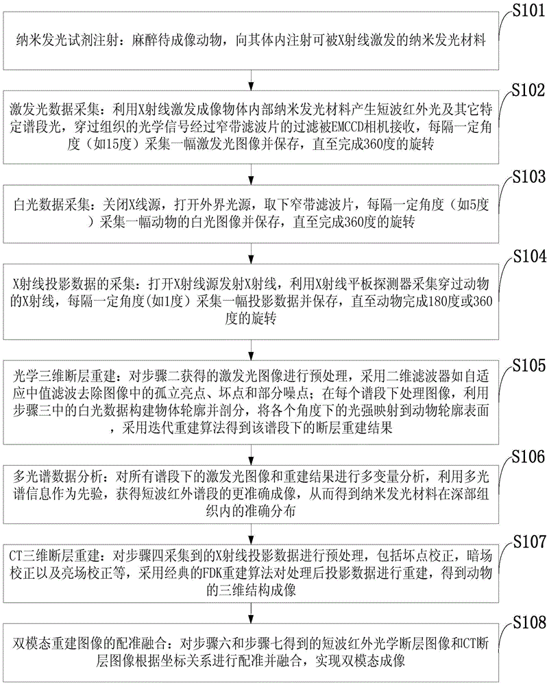
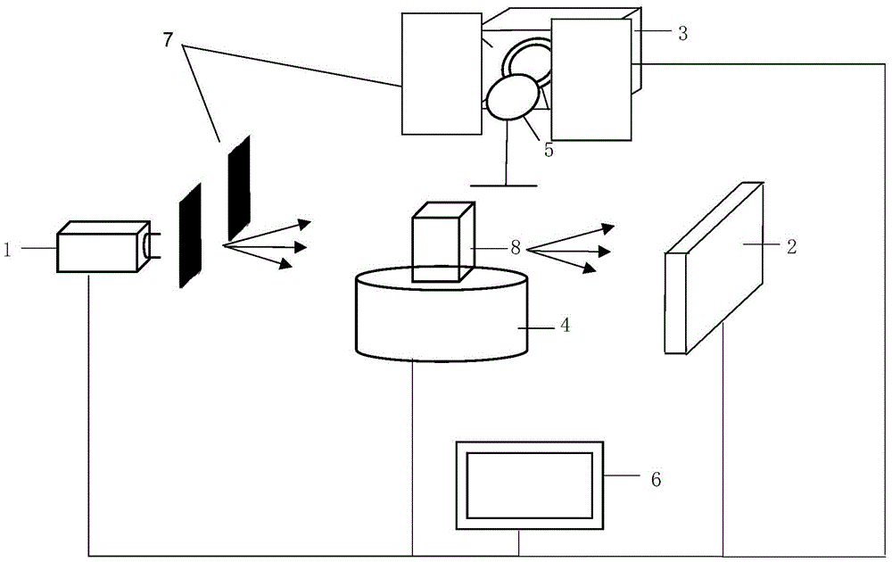
[0039] Such as figure 1 Shown: A method for X-ray-excited multi-spectral tomography in deep tissue, which uses X-rays to excite nano-luminescent materials to emit specific spectrum bands, especially short-wave infrared light with a higher penetration depth, and the photons pass through the tissue to reach the imaging object On the surface, after being filtered by a narrow-band filter, the light of different wavelength bands is received by the EMCCD camera, and the imaging and reconstruc...


PUM


Abstract

The invention discloses a deep tissue X-ray excitation multispectral tomography system and method. The system comprises an X-ray source, an X-ray flat panel detector, an EMCCD camera, an electric control rotary table, a narrow-band filter, a lead plate, a computer and an animal to be imaged; nano-luminescence materials are excited through an X ray, short-wave infrared light with the specific spectra sections, particularly high penetration depth is emitted, photons penetrate through tissue to reach the surface of an imaged object, the light of different wave length spectra sections is received by an EMCCD camera by being filtered by the narrow-band filter, and an obtained image and a rebuilt result under each spectra section are obtained; multivariate analysis is adopted for carrying out treatment and analysis on multispectral images and rebuilt results, accurate distribution of the nano-luminescence materials in the object is obtained and combined with CT tomography results, and finally a deep tissue multispectral tomography image is obtained. The imaging depth of X-ray excitation multispectral tomography can be effectively improved, and deep tissue imaging of small living animals is achieved.

Description

technical field [0001] The invention belongs to the technical field of multi-modal molecular imaging, and in particular relates to a method and system for deep tissue X-ray excitation multi-spectral tomography. Background technique [0002] Optical molecular imaging technology can observe the changes of molecular levels in living organisms in vivo, non-contact and dynamically, and provides effective technical means for solving some important problems in the field of biomedical research. However, the strong scattering characteristics of light in biological tissues greatly limit the penetration depth and imaging resolution of optical imaging, and also limit the application of optical imaging technology in deep tissues and organs. [0003] X-ray luminescence computed tomography (XLCT) is a new multi-modal molecular imaging technology that combines CT tomography and optical molecular imaging. It was first proposed in 2010 by the research group of Xing Lei from Stanford Universit...

Claims


Application Information

Patent Timeline
IPC IPC(8): A61B6/03A61B6/10
CPCA61B6/035A61B6/107A61B6/4417A61B6/482A61B6/508A61B6/5211A61B6/5247
Inventor 卢虹冰高鹏戎军艳刘文磊廖琪梅刘洋常小红
Owner FOURTH MILITARY MEDICAL UNIVERSITY
Why Eureka
Try Eureka