Sample preparation method employing AFM (Atomic Force Microscope) for single antibody molecule imaging

An atomic force microscope and antibody molecule technology, applied in the nanometer field, can solve the problems of unsatisfactory imaging effect of single antibody molecule IgG and complicated preparation methods

Active Publication Date: 2016-06-22
SHANGHAI INST OF APPLIED PHYSICS - CHINESE ACAD OF SCI
View PDF5 Cites 8 Cited by
  • Summary
  • Abstract
  • Description
  • Claims
  • Application Information

AI Technical Summary

Problems solved by technology

[0004] The technical problem to be solved by the present invention is to overcome the problems in the prior art that the sample preparation method for atomic force microscope imaging of a single antibody molecule is re

Method used


Image

  • Sample preparation method employing AFM (Atomic Force Microscope) for single antibody molecule imaging
  • Sample preparation method employing AFM (Atomic Force Microscope) for single antibody molecule imaging
  • Sample preparation method employing AFM (Atomic Force Microscope) for single antibody molecule imaging

Examples


Example Embodiment

[0048] Example 1 Sample preparation for AFM imaging of a single digoxin antibody molecule IgG
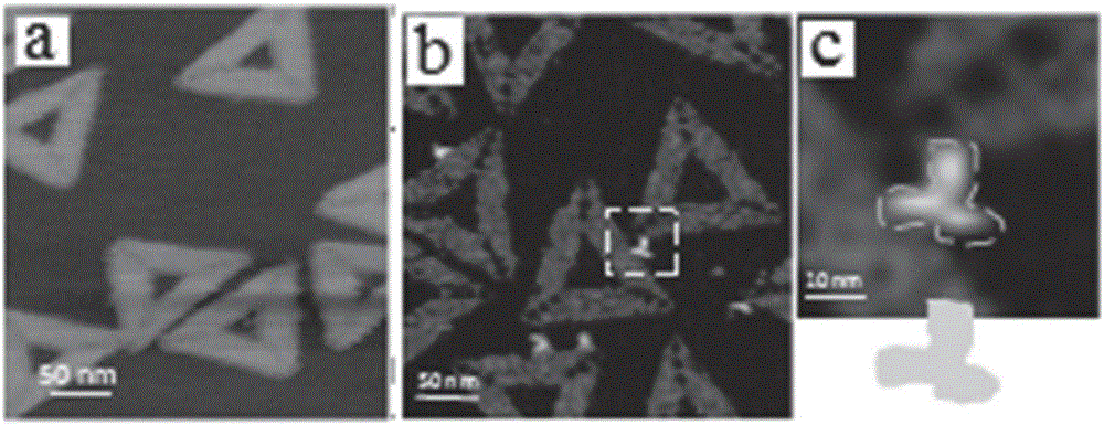
[0049] 1) Mix the single-stranded scaffold strand DNAM13mp18 and the staple strand DNA coupled with digoxin in the TAE-Mg at a molar ratio of 1:10 2+ Buffer system (40mMTris-acetic acid, 1mMEDTA, 12.5mMMgCl 2 , PH8.0), annealed from 95°C to 20°C with a PCR machine, and an annealing rate of 0.1°C / 10s, to prepare an equilateral triangle DNA origami coupled with digoxin; the side length of the equilateral triangle DNA origami The diameter is 130 nm, and there are 3 digoxin antigens with equal spacing on each side, and each digoxin antigen contains 2 digoxin molecules coupled to DNA origami with a distance of about 10-15 nm between each other; The nucleotide sequence of the staple strand DNA coupled with digoxin was published on March 17, 2010 in J.Am.Chem.Soc.Volume 132, Issue 10, Page 3248-3249, entitled Goldnanoparticleself- The DNA sequence from A01 to Loop described on pages S11-S16 i...

Example Embodiment

[0053] Example 2 Sample preparation for AFM imaging of a single digoxigenin antibody molecule
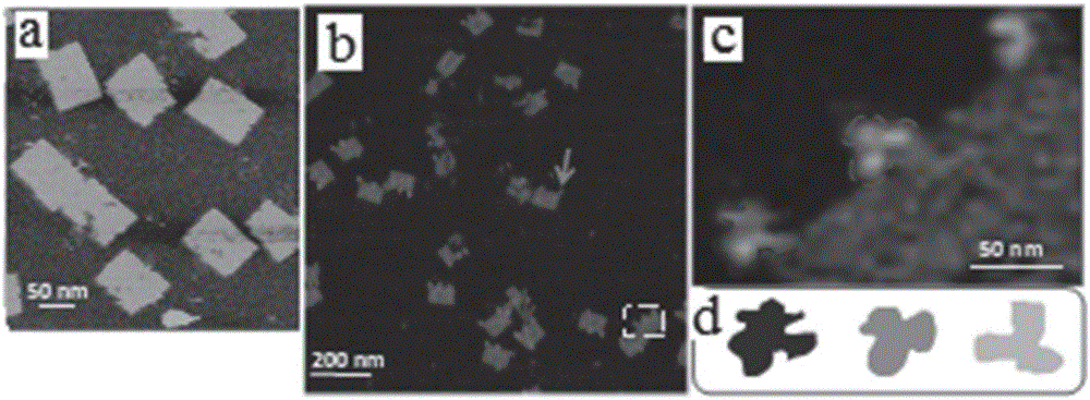
[0054] 1) Mix the single-stranded scaffold strand DNAM13mp18 and the staple strand DNA coupled with digoxin in the TAE-Mg at a molar ratio of 1:10 2+ Buffer system, using PCR machine to anneal from 95°C to 20°C, with an annealing rate of 0.1°C / 10s, to prepare a rectangular DNA origami coupled with digoxin; the side length of the rectangular DNA origami is 100nm×70nm. There are 3 digoxin antigens with equal spacing on each side, and each digoxin antigen contains 2 digoxin molecules coupled to DNA origami with a distance of about 8-13nm between each other; the coupling The nucleotide sequence of the staple strand DNA with digoxin is in the paper titled Self-AssembledWater-SolubleNucleicAcidProbeTilesforLabel-FreeRNAHybridizationAssays published on pages 180-183 of Science Volume 319, Issue 5860 on January 11, 2008 The DNA sequence from 1 to 216 described on pages S15-S19 of the supplemen...

Example Embodiment

[0058] Example 3 AFM imaging of a single digoxin antibody molecule IgG

[0059] The AFM imaging of the single antibody molecule IgG prepared in Example 1 was subjected to AFM scanning imaging using a "J" scanning head. The AFM probe was Bruke's silicon nitride probe (SNL, elastic coefficient 0.35N / m). The AFM imaging mode is the liquid phase "tap" mode, select the local scanning range, the imaging temperature is controlled at about 25℃, the humidity is controlled at about 40%, the scan rate is controlled below 2Hz, and the scan parameters are adjusted in the low force area (F< 200pN), the scan time is 1 minute.

[0060] Scan imaging results such as figure 1 As shown, the DNA origami pattern of equilateral triangle coupled with digoxin adsorbed on the surface of mica is complete and clear, such as figure 1 Shown in a and 1b. The length of the side of the equilateral triangle is 130nm. There are five antibody molecules IgG observed on the equilateral triangle DNA origami. Two of t...


PUM

PropertyMeasurementUnit
Scaleaaaaaaaaaa

Abstract

The invention discloses a sample preparation method employing an AFM (Atomic Force Microscope) for single antibody molecule imaging, a single antibody molecule IgG AFM imaging method and a DNA origami combined with antibody molecule IgG. The preparation method includes (1) mixing and reacting the DNA origami coupled with micromoleculeantigen at the edge with antibody of the micromoleculeantigen, wherein the micromoleculeantigen includes two identical micro molecules coupled to the edge of the DNA origami and spaced from each other by about 8-15 nanometers; (2) dropwise adding a reaction product on a newly-dissolved mica plate for adsorbing and imaging, and adding a buffer solution. Therefore, a sample is obtained. The preparation method provided by the invention is simple and convenient in operation. The antibody molecule IgG is absorbed on the mica surface with the aid of the DNA origami in a liquid environment, so that solid liquid interphasecapillary force effect on the antibody molecule IgG can be avoided and influence of a pinpoint effect on the antibody molecule IgG structure is reduced; and the imaging effect is improved.

Description

technical field [0001] The invention relates to the field of nanotechnology, in particular to a sample preparation method for single antibody molecule atomic force microscope imaging, an atomic force microscope imaging method for single antibody molecule IgG and a DNA origami combined with antibody molecule IgG. Background technique [0002] Atomic force microscopy (AFM) is one of the important tools for studying biomolecules at the nanoscale. Because of its ability to image biomolecules at the nanometer level under near-physiological conditions and obtain high-definition molecular morphology features, it has become an irreplaceable research method for studying the structure and function of biomolecules. [0003] Antibodies are an important class of biomolecules. They can neutralize toxins and viruses, promote phagocytosis by phagocytic cells, mediate immune cell activity, and stimulate complement, which play a very important role in the prevention, diagnosis, and treatment ...

Claims


Application Information

Patent Timeline
IPC IPC(8): G01Q30/20
CPCG01Q30/20
Inventor 周星飞赵志杰张萍李宾虞国凯胡钧郝长春杨家香
Owner SHANGHAI INST OF APPLIED PHYSICS - CHINESE ACAD OF SCI
Why Eureka
Try Eureka