
Measuring method for cell potential of bioengineering retina nerve scaffold

A technology of bioengineering and measurement methods, applied in the field of stem cell tissue engineering and neurobiology, can solve the problems of low cell survival rate, incomplete growth and differentiation of transplanted cells, integration of host cells, etc.

Inactive Publication Date: 2016-07-06
ZHONGSHAN OPHTHALMIC CENT SUN YAT SEN UNIV
View PDF6 Cites 1 Cited by
  • Summary
  • Abstract
  • Description
  • Claims
  • Application Information

AI Technical Summary

Problems solved by technology

However, there are many complex and critical problems in stem cell transplantation: for example, it is difficult to precisely control the cell growth site; the transplanted cells cannot fully grow and differentiate, and it is difficult to effectively integrate with the host cells to form functional synaptic links; the survival rate of cells transplanted into the eye is low and many more

Method used


Image

  • Measuring method for cell potential of bioengineering retina nerve scaffold
  • Measuring method for cell potential of bioengineering retina nerve scaffold
  • Measuring method for cell potential of bioengineering retina nerve scaffold

Examples


Embodiment 1

[0019] 1. Production of bioengineered retinal scaffold (iRP):

[0020] 1. Using the method of mechanical separation, put the nerve fiber layer of human iPSc-derived 3D retina cultured for 50-60 days (specific preparation method reference: XiufengZhong, et al. Generation of three-dimensional retinal tissue with functional photoreceptors from human iPSCs, NatCommun.2014Jun10; 5:4047) Put it into D-Hanks solution, use a 0.45mm needle to separate the tissue under a 50 times upright microscope, discard the pigmented part (pigment layer) in the retinal tissue, and keep the golden yellow tissue part (nerve fiber layer); the separated nerve fiber Use a pipette to transfer the layered tissue to a 3.5cm culture dish, rinse with PBS for 10 minutes; absorb the PBS, digest the cells with accutase cell digestion solution, and place them in a 37°C incubator for 30 minutes; at the end of the process, the cells can be sucked into the centrifuge tube and blown. Under a 10x upright microscope, i...


PUM


Abstract

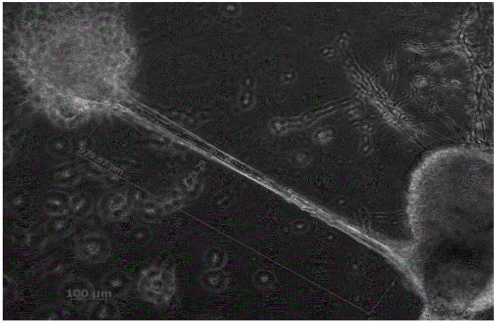
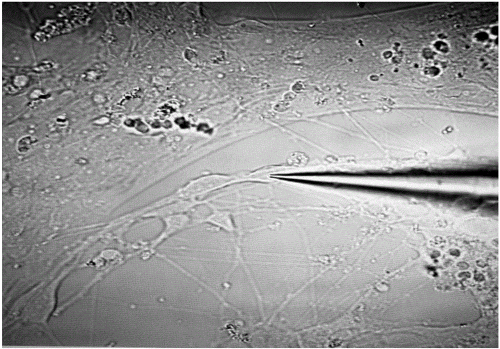
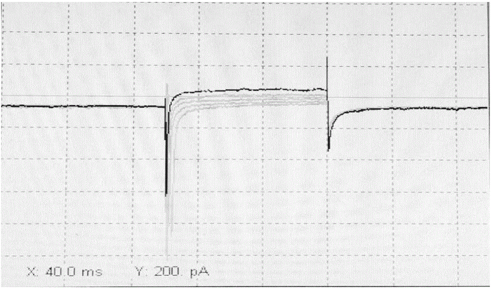
The invention discloses a measuring method for cell potential of a bioengineering retina nerve scaffold. The measuring method includes the steps that a nerve fiber layer of human iPSc-derived 3D retina is digested into unicells, the unicells serving as seed cells are resuspended through an induced differentiation medium, then polylactic acid-glycolic acid polymer support coated with matrigel Matrigel is inoculated with the unicells, induced differentiation is conducted in the induced differentiation medium to form the bioengineering retina nerve scaffold, wherein the induced differentiation medium is a medium containing neurotrophic factors and a retina differentiation nutrient solution for maintaining the concentration; ion channel current recording is conducted on nerve-like cells, cultured to a certain stage, on the bioengineering retina nerve scaffold in a whole-cell recording mode, and the neural basis function of the bioengineering retina nerve scaffold is reflected through potential detection of the scaffold cells.

Description

Technical field: [0001] The invention belongs to the fields of stem cell tissue engineering and neurobiology, and in particular relates to a method for measuring the potential of bioengineered retinal nerve scaffold cells. Background technique: [0002] Glaucoma is a group of clinical syndromes or eye diseases that threaten and damage the optic nerve and its visual pathways, eventually leading to impairment of visual function, mainly related to pathological elevated intraocular pressure. Its optic neuropathy is characterized by progressive loss of retinal ganglion cells (RGCs) and damage to their axonal connections to the central brain. At present, the main treatment for lowering intraocular pressure clinically can slow down the progression of the disease, but it cannot completely prevent the further development of the disease, especially for patients with advanced glaucoma. Therefore, it is necessary to find a new therapeutic strategy to slow down or stop the progressive R...

Claims


Application Information

Patent Timeline
Patent Type & Authority Applications(China)
IPC IPC(8): G01N27/60
CPCG01N27/60
Inventor 葛坚顾怀宇李康寯
Owner ZHONGSHAN OPHTHALMIC CENT SUN YAT SEN UNIV
Learn More