Nano-gold CT contrast agent for early diagnosis of hepatocellular carcinoma and preparation method of contrast agent
An early diagnosis and nano-gold technology, applied in the field of biomedicine, can solve the problems of unsuitable animal CT experiments and poor stability, and achieve the effects of improving enrichment and tissue penetration, improving stability, and good biocompatibility
- Summary
- Abstract
- Description
- Claims
- Application Information
AI Technical Summary
Problems solved by technology
Method used
Image
Examples
Embodiment 1
[0045] Example 1: Preparation of nano-gold CT contrast agent for early diagnosis of liver cancer
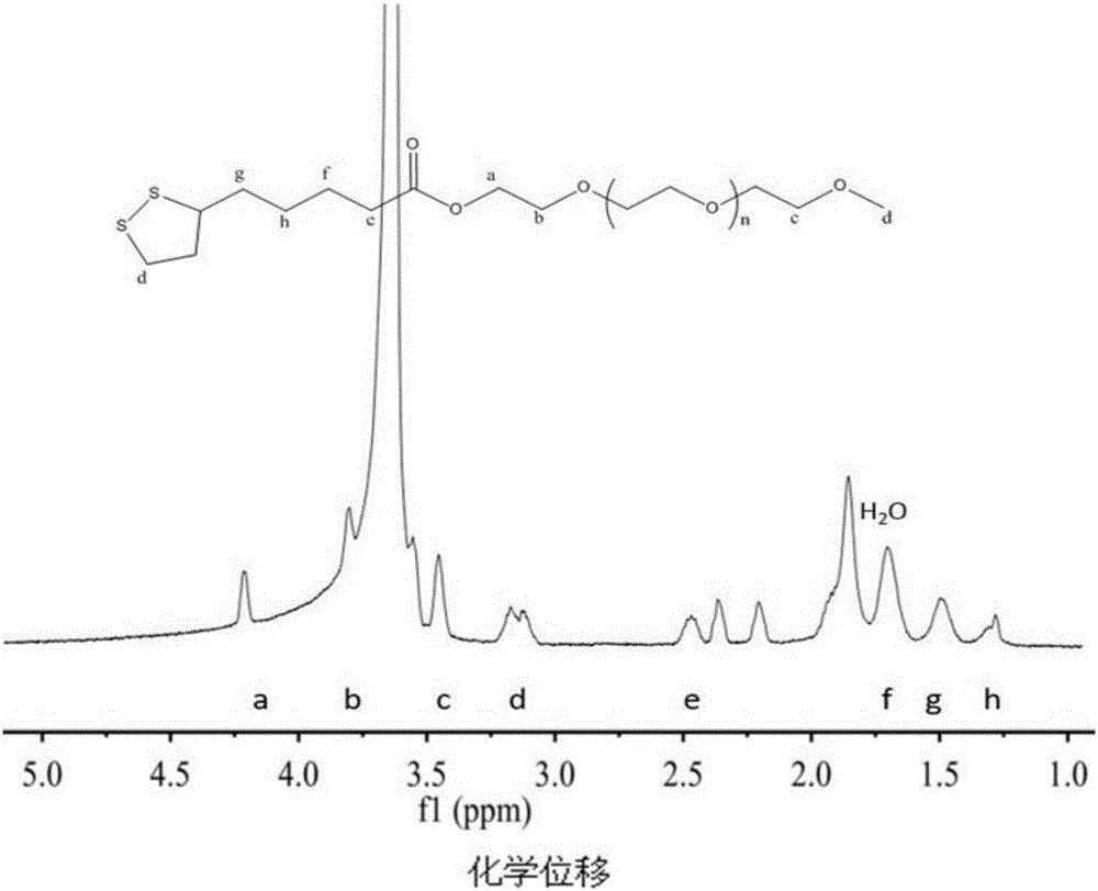
[0046] 1) LA-PEG 2k Synthesis: Weigh 2 g of single-ended methoxy mPEG 2k -OH and 0.206g lipoic acid (LA) (the molar ratio is LA:mPEG 2k -OH=5:1) was added to a 50mL round bottom flask, and 960mg of 1-ethyl-(3-dimethylaminopropyl)carbodiimide hydrochloride (EDC.HCL) and 610mg of 4-bis Dimethylaminopyridine (DMAP), dissolved in 30 mL of dichloromethane (DCM). Stir in an ice-water bath for 30 minutes, then react in a water bath at 35°C for 2 days. After the reaction, dichloromethane was removed by rotary evaporation. The remaining solid was dissolved in distilled water, transferred to a dialysis bag (MWCO 2k) and dialyzed for two days. lyophilized to obtain LA-PEG 2k White powdery solid. Characterized by NMR, see attached image 3 .
[0047] 2) LA-PEG 4k and LA-PEG 10k The synthesis of: method is the same as step 1.
[0048] 3) Preparation of AuNPs: Weigh 59 mg tetrachlo...
Embodiment 2
[0058] Example 2: Nano-gold CT contrast agent ligand shielding-deshielding investigation:
[0059] 1) Assembly-disassembly investigation of nano-gold CT contrast agent
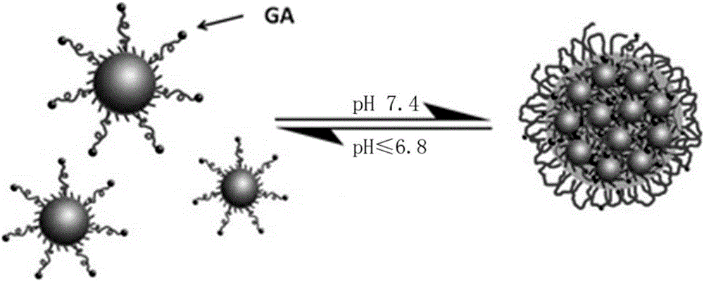
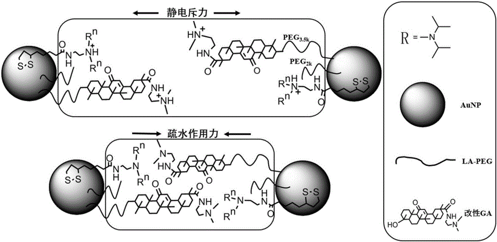
[0060] For nano gold CT contrast agent, its assembly-disassembly properties under normal tissue (pH7.4) and tumor conditions (pH6.8) were investigated. In the experimental group 1:4 (see attached figure 2 ) and control group 1 as an example, the details are as follows: Add NaOH solution (pH13) dropwise to the nano-gold solution to pH8-9. Solutions HEPESpH7.4 and HEPESpH6.8, use dynamic light scattering (DLS) to investigate the particle size at different pHs, the particle size of the assembly is 100-200nm, and the particle size of the disassembly is 30-40nm, which is used to illustrate the assembly-disassembly of the system nature. Then the pH of the solution was adjusted repeatedly between pH 7.4 and pH 6.8, and the particle size change of the test system was used to investigate the pH-sensitive reversible...
Embodiment 3
[0066] Example 3: Investigation on Concentrated Stability of Nano-gold CT Contrast Agent:
[0067] Batch centrifugation is used to concentrate the modified nano-gold solution (ie nano-gold CT contrast agent). For the first centrifugation (25,000 rpm, 45 minutes), the batch of nano-gold liquid is concentrated about 20 times; for the second centrifugation (15,000 rpm, 60 minutes), the concentrated solution obtained by the first centrifugation is further concentrated about 8 times. Finally, the nano-gold concentrated solution (0.15M) with a concentration (0.1M) suitable for CT imaging of tumor-bearing mice was obtained. Precipitation occurred in the experimental group 1:0 and the experimental group 1:2, which indicated that the contrast agent had poor stability at the contrast concentration and was not suitable for in vivo CT imaging. The test results are attached Figure 10 .
[0068] Combining Example 2 and Example 3, it is confirmed that the gold nano CT contrast agent suit...
PUM
 Abstract
Description
Claims
Application Information
 - R&D
- Intellectual Property
- Life Sciences
- Materials
- Tech Scout
- Unparalleled Data Quality
- Higher Quality Content
- 60% Fewer Hallucinations
Browse by: Latest US Patents, China's latest patents, Technical Efficacy Thesaurus, Application Domain, Technology Topic, Popular Technical Reports.
© 2025 PatSnap. All rights reserved.Legal|Privacy policy|Modern Slavery Act Transparency Statement|Sitemap|About US| Contact US: help@patsnap.com


