
Corneal limbal stem cell cryopreservation liquid and cryopreservation method

A technology of corneal limbal stem cells and cryopreservation method, which is applied in the direction of non-embryonic pluripotent stem cells, animal cells, vertebrate cells, etc., and can solve the problems of damaging the biological characteristics and damage of corneal limbal stem cells

Active Publication Date: 2016-08-17
GUANGZHOU SALIAI STEMCELL SCI & TECH CO LTD
View PDF5 Cites 8 Cited by
  • Summary
  • Abstract
  • Description
  • Claims
  • Application Information

AI Technical Summary

Problems solved by technology

However, studies have shown that the existing low-temperature freezing scheme has greatly damaged the biological characteristics of corneal limbal stem cells, and the damage will become more obvious as time goes by

Method used


Image

  • Corneal limbal stem cell cryopreservation liquid and cryopreservation method
  • Corneal limbal stem cell cryopreservation liquid and cryopreservation method
  • Corneal limbal stem cell cryopreservation liquid and cryopreservation method

Examples

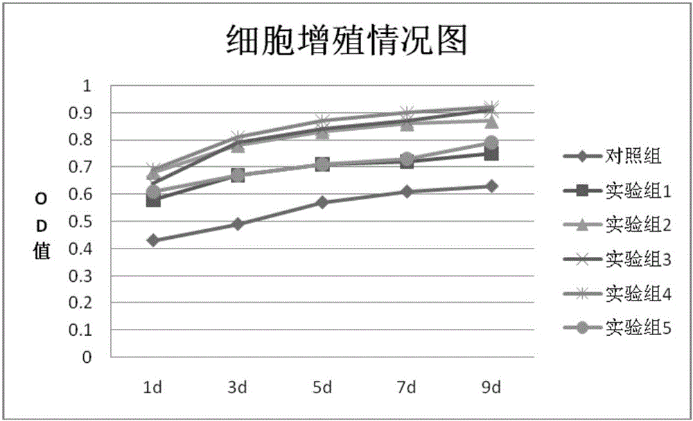
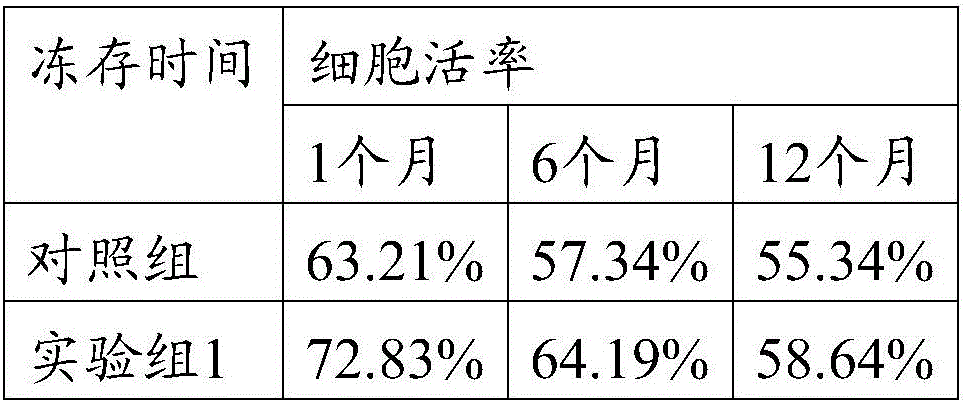

Embodiment 1

[0033] Embodiment 1, the cryopreservation solution of limbal stem cells

[0034] The specific ingredients are as follows:

[0035]

Embodiment 2

[0036] Example 2, Isolation and Culture of Rabbit Limbal Stem Cells

[0037] 1. Take fresh eyeballs from rabbits in a sterile environment.

[0038] 2. In the ultra-clean bench, cut off the conjunctival tissue, rinse with normal saline, soak in normal saline containing double antibodies for 30 minutes, cut off the cornea along the edge of the cornea, place it in double-antibody (penicillin and streptomycin), and dissect it under a dissecting microscope. Next, remove the iris tissue, drill the central cornea with an 8.5 trephine drill, and cut the corneal ring and central corneal tissue into pieces.

[0039] 3. According to the volume of the tissue block, every 1cm 2 Add 1 mL of 1% collagenase + 0.5% neutral protease to the tissue block, digest at 37°C for 40-60 min, add PBS containing 10% FBS to each 1 mL of digestion solution to stop digestion, filter with 100 μm mesh, centrifuge at 1000 rpm for 5 min, discard the supernatant, Add (DMEM+10%FBS) medium to 1×10 5 / mL density ...

Embodiment 3

[0040] Embodiment 3, the cryopreservation scheme of limbal stem cells

[0041] 1. When the confluence of the bottom layer of each cell culture dish reaches 80%, remove the supernatant culture solution on the culture dish, add 0.25% trypsin, digest for 5-10min, add PBS containing 10% FBS to every 1mL digestion solution to stop digestion, Centrifuge at 1000rpm for 5min, remove the supernatant, and add freezing solution.

[0042] 2. Put it into the program cooling device to cool down, design the cooling program as follows: normal temperature to 4°C, drop 5°C per minute; 4°C to -10°C, drop 1°C per minute; -10°C to -80°C, drop every minute 3°C, -80°C to -196°C, drop 5°C per minute.

[0043] 3. After the program cools down to -196°C, store it in liquid nitrogen.


PUM


Abstract

The invention relates to the field of stem cells and discloses corneal limbal stem cell cryopreservation liquid and cryopreservation method. The corneal limbal stem cell cryopreservation liquid comprises CHIR99021. Compared with conventional corneal limbal stem cell cryopreservation liquid, after cryopreserved with the corneal limbal stem cell cryopreservation liquid, stem cells recover, cell viability is obviously higher than that of cells cryopreserved with other types of conventional cryopreservation liquid, the cell viability capability is also superior to that of cells cryopreserved with conventional cryopreservation liquid, and the corneal limbal stem cell cryopreservation liquid can be used for long-time preservation and application of the corneal limbal stem cells. The corneal limbal stem cell cryopreservation method comprises the steps of conducting digestion and centrifugation on the corneal limbal stem cells, then removing supernatant, adding the corneal limbal stem cell cryopreservation liquid, and sub-packing the corneal limbal stem cells in cryopreservation tubes for cryopreservation. Compared with conventional cryopreservation methods, after the cells cryopreserved through the corneal limbal stem cell cryopreservation method recover, cell viability is obviously higher than that of cells cryopreserved through other types of conventional cryopreservation methods, and the cell viability capability is also superior to that of cells cryopreserved thorugh conventional cryopreservation methods.

Description

technical field [0001] The invention relates to the field of stem cells, in particular to a cryopreservation solution and a cryopreservation method for limbal stem cells. Background technique [0002] The corneal limbus is the junction of the cornea, conjunctiva and sclera. The distinguishing mark from the cornea is the termination of Bowman's membrane; the distinguishing mark from the conjunctiva is that it does not contain goblet cells and is about 1 mm wide. There are only epithelial and stroma layers here. The epithelial cell layer is more than 10 layers, arranged irregularly, the cells are small cylindrical, and the nucleus is deeply stained. The deep basal cells are a layer of small cylindrical or cuboid cells, and the nuclei are oval and parallel to the surface. In the basal The papillae are formed in the papilla, forming a special "palisade"-like epithelial structure, which contains pigment and rich vascular network, and is closely connected with the basement membran...

Claims


Application Information

Patent Timeline
Patent Type & Authority Applications(China)
IPC IPC(8): A01N1/02C12N5/074
CPCA01N1/0221C12N5/0602
Inventor 葛啸虎陈海佳王一飞吴子杰
Owner GUANGZHOU SALIAI STEMCELL SCI & TECH CO LTD
Learn More