
Fluorescent microsphere supported optical material and preparation method thereof

A technology of fluorescent microspheres and optical materials, applied in the field of optical testing materials, can solve the problem that biochips are not mentioned in relevant literature, and achieve the effect of significant reference significance, suitable spacing, and correcting imaging quality.

Active Publication Date: 2016-08-17
MGI TECH CO LTD
View PDF15 Cites 0 Cited by
  • Summary
  • Abstract
  • Description
  • Claims
  • Application Information

AI Technical Summary

Problems solved by technology

FluoSpheres are mostly used in the biomedical field, but there is no relevant literature mentioning their application to the fluorescence test of biochips

Method used


Image

  • Fluorescent microsphere supported optical material and preparation method thereof
  • Fluorescent microsphere supported optical material and preparation method thereof
  • Fluorescent microsphere supported optical material and preparation method thereof

Examples


Embodiment 1

[0034] Embodiment 1, preparation of optical material loaded with fluorescent microspheres

[0035] (1) Rinse the silicon-based chip with 50 μL of 50 mM phosphate buffer solution and 50 μL of 50 mM sodium hydroxide aqueous solution in sequence, incubate for 20 min while rinsing with aqueous sodium hydroxide solution, and then wash the silicon-based chip with 50 μL of 50 mM phosphate buffer solution to make the silicon-based chip There are positively charged amino groups on the surface of the chip;

[0036] (2) 2-(N-morpholine) ethanesulfonic acid solution of 3 mg / mL 1-(3-dimethylaminopropyl)-3-ethylcarbodiimide with a volume ratio of 10:1 and Carboxylated fluorescent microspheres (F-8809, with a wavelength of 530nm and a diameter of 0.2μm) were mixed uniformly by shaking for 5s and centrifuging for 5s in turn to obtain a mixture;

[0037] (3) Rinse the silicon-based chip treated in step (1) with the mixed solution, bond at 21° C. for 30 min, and rinse with 50 mM phosphate buff...

Embodiment 2

[0044] Embodiment 2, preparation of optical material loaded with fluorescent microspheres

[0045] The preparation method is the same as that in Example 1, except that the silicon-based chip is replaced by a glass chip (ordinary glass sheet).

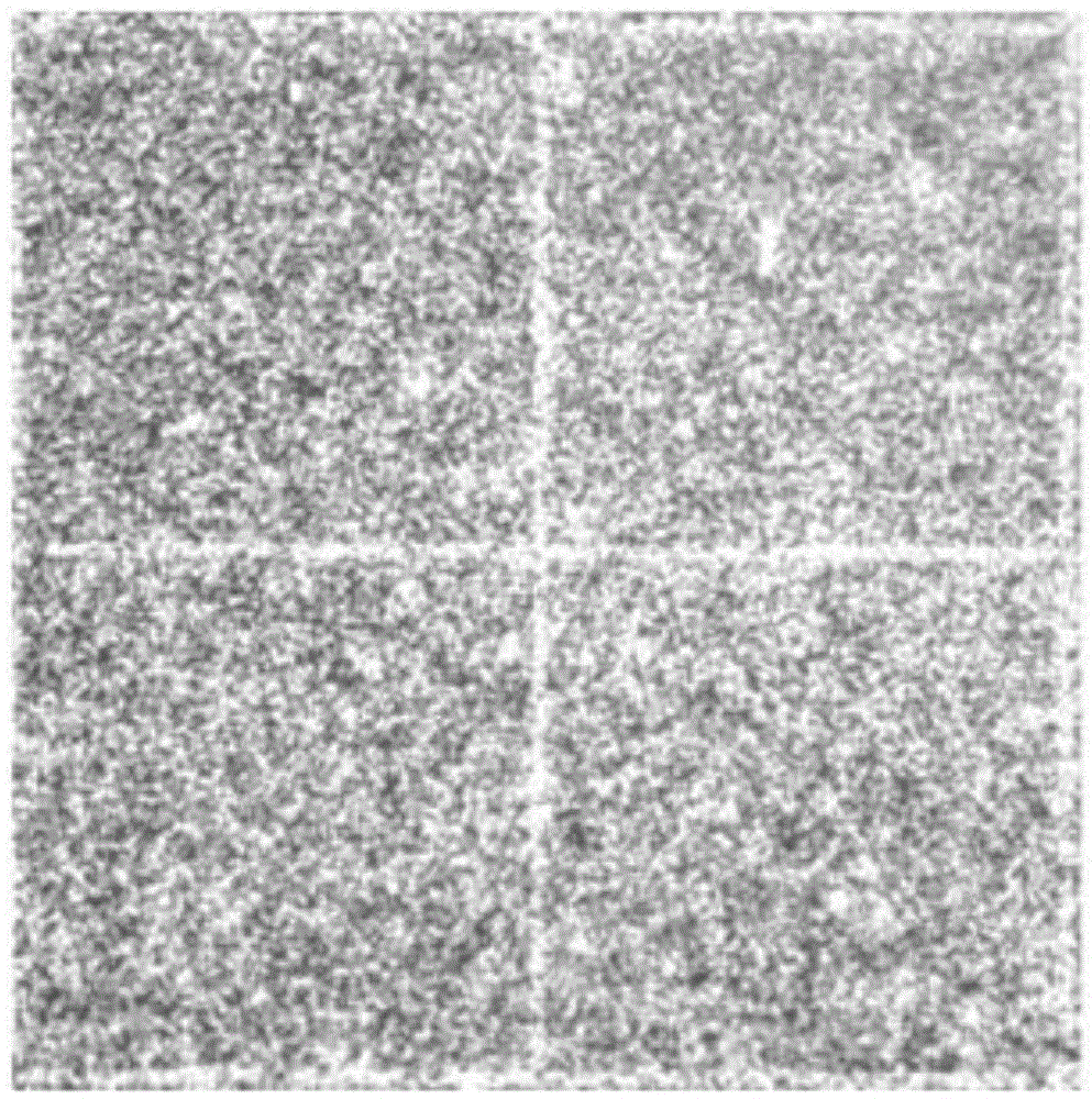
[0046] In this embodiment, the optical material loaded with fluorescent microspheres is exposed under a 40 times optical microscope through a green excitation light for 50 ms, and the optical material loaded with fluorescent microspheres can be seen as bright spots under the optical microscope, which is bonding on the surface of the glass chip. The fluorescence emitted by the fluorescent microspheres under the corresponding wavelength of 530nm excitation proves that the distance between the fluorescent microspheres bonded to the surface of the glass chip is suitable and even.


PUM

PropertyMeasurementUnit
diameteraaaaaaaaaa
diameteraaaaaaaaaa

Abstract

The present invention discloses a preparation method of a fluorescent microsphere supported optical material. The method comprises the steps of: (1) sequentially flushing a biochip with phosphate buffer, sodium hydroxide aqueous solution and a phosphate buffer solution; (2) mixing 2-(N-morpholino) ethanesulfonic acid solution of 1-(3-dimethylaminopropyl)-3-ethyl carbodiimide imine with carboxylated fluorescent microspheres to obtain a mixed solution; and (3) rinsing the biochip treated in the step (1) with the mixed solution, and bonding to obtain the optical material. The invention has the following advantages: carboxylated fluorescent microspheres are smoothly and uniformly bonded to the surface of the biochip at suitable pitch, and can be used to test autofocus system, lasers and filters; the fluorescing time is up to half an hour, which is enough for operators to debug the optical system, so as to help correct the image quality of the optical imaging system. The optical material has size, shape and bonding mode very similar with the actual substance for sequencing, and has significant reference value for debugging.

Description

technical field [0001] The invention belongs to the field of optical testing materials for sequencers, and in particular relates to an optical material loaded with fluorescent microspheres and a preparation method thereof. Background technique [0002] In the process of debugging the optical system, a test chip is usually used to adjust the focal plane and test the laser. In the prior art, the method of coating fluorescence on the silicon-based chip retrieves the LED chip and mixes the phosphor powder and the glue by dispensing glue, and points it on the surface of the silicon-based chip (a method of spotting fluorescent glue on LED based on COB technology) , publication date 2011.08.10, publication number CN102148299A). If the same method is adopted and the fluorescent microspheres and glue are mixed and spotted on the surface of the silicon wafer, the fluorescence cannot be guaranteed to be on the same plane and its uniformity. [0003] Fluorescent microspheres ( Fluor...

Claims


Application Information

Patent Timeline
Patent Type & Authority Applications(China)
IPC IPC(8): G01N21/64
Inventor 冯赫天张永卫倪鸣代冲冲
Owner MGI TECH CO LTD
Learn More