
Cup-type time-resolved fluorescent immunoassay kit for high-sensitivity C-reactive protein based on microspheres, and preparation method and application thereof

A time-resolved fluorescence and protein-reactive technology, which is applied in the field of clinical medical diagnosis, can solve the problems of difficult routine implementation in clinical laboratories, unsatisfactory sensitivity and specificity, and high technical requirements for chemiluminescence, so as to achieve easy automatic operation and shorten detection time , The effect of improving the detection sensitivity

Inactive Publication Date: 2016-08-31
SHANGHAI UPPER BIO TECH PHARMA
View PDF2 Cites 3 Cited by
  • Summary
  • Abstract
  • Description
  • Claims
  • Application Information

AI Technical Summary

Problems solved by technology

Latex turbidimetry generally uses polyclonal antibodies to coat latex microspheres, and the sensitivity and specificity are not ideal. If monoclonal antibodies are used to coat latex microspheres, the cost of reagents is very high
The chemiluminescence method has high technical requirements, and the operation steps are relatively cumbersome. It requires a multi-step reagent addition process, and it is not easy to carry out routinely in clinical laboratories.
Although the colloidal gold immunochromatography method has the advantages of less sample consumption, simple, quick, and cheap, however, when the antigen or antibody content in some samples is extremely low, the color of the colloidal gold will be very light or even no color, and it is difficult to Judging the results with the naked eye is prone to misjudgment and low sensitivity
Although the sensitivity of fluorescence immunochromatography is higher than that of colloidal gold immunochromatography, its detection precision is not ideal, and the sensitivity is still far behind that of large-scale chemiluminescence or electrochemiluminescence instruments

Method used

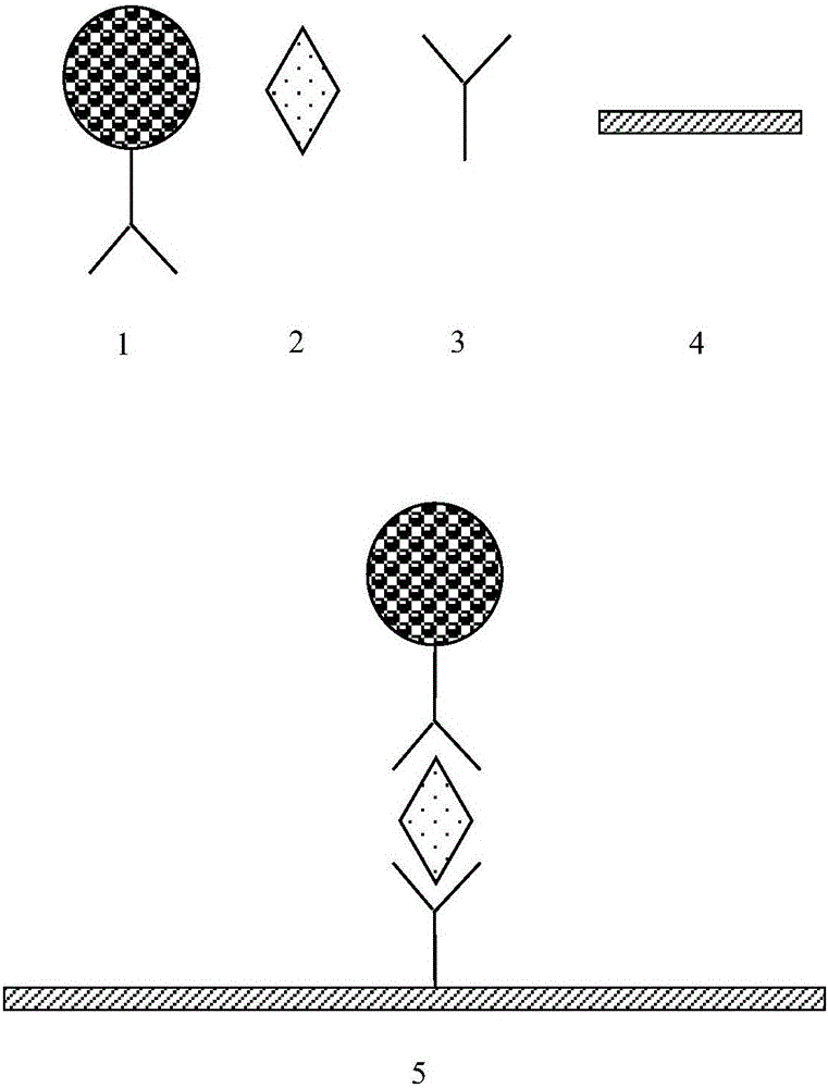

Image

  • Cup-type time-resolved fluorescent immunoassay kit for high-sensitivity C-reactive protein based on microspheres, and preparation method and application thereof
  • Cup-type time-resolved fluorescent immunoassay kit for high-sensitivity C-reactive protein based on microspheres, and preparation method and application thereof
  • Cup-type time-resolved fluorescent immunoassay kit for high-sensitivity C-reactive protein based on microspheres, and preparation method and application thereof

Examples


Embodiment 1

[0044] (1) Preparation of detection cuvette:

[0045] A. Detection cuvette coating: C-reactive protein antibody 6404 (Medix Company) was diluted to 10 μg / ml with 0.2mol / L phosphate buffer (pH7.8), 100 μL / well, coated at 37°C for two hours, and washed. .

[0046] B. Detection cuvette blocking: Add 200 μL / well of blocking buffer to the cuvette, block at 37°C for two hours, discard the blocking solution in the coated cuvette, and pat dry.

[0047] C. Drying of the test cuvette: place the sealed cuvette above in a 37° C. drying oven with a humidity lower than 30% for 4 hours, and store in a sealed and dry place.

[0048] (2) Preparation of fluorescently labeled antibodies:

[0049] A. Fluorescent particle activation:

[0050] Take 1mg of carboxyl time-resolved fluorescent microspheres (300nm, 0.1ml, 10mg / mL, Bangslab Company), add 60μL of 500mmol / L MES (2-(N-morpholine)ethanesulfonic acid) buffer, pH 6.0, add Add purified water to 0.2mg 1-(3-dimethylaminopropyl)-3-ethylcarbodi...

Embodiment 2

[0060] (1) Drawing of standard curve:

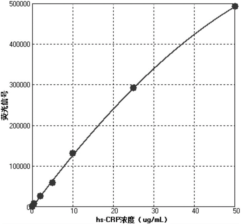
[0061] The reagents prepared in Example 1 were combined into a high-sensitivity C-reactive protein time-resolved fluorescence quantitative kit, and the calibrator was measured, and each concentration was repeated 10 times.

[0062] Add 3 μL of calibrator and 100 μL of fluorescently labeled antibody to each detection, incubate at 37°C for 20 min, then wash the detection cup, and read on the Victor X4 fluorescence reader from PerkinElmer (excitation wavelength 340-380nm, detection wavelength 600-630nm) The specific data are shown in Table 1.

[0063] Table 1

[0064]

[0065] According to the data in Table 1, the concentration of the calibrator is taken as the abscissa, and the mean fluorescence signal is taken as the ordinate to draw a standard curve. standard curve as figure 2 shown. The standard curve has good linearity, and the concentration of high-sensitive C-reactive protein contained in the sample can be quantitatively anal...

Embodiment 3

[0073] (1) Preparation of detection cuvette:

[0074] A. Detection cuvette coating: C-reactive protein antibody 6404 (Medix Company) was diluted to 10 μg / ml with 0.2mol / L phosphate buffer (pH7.8), 100 μL / well, coated at 37°C for two hours, and washed. .

[0075] B. Detection cuvette blocking: Add 200 μL / well of blocking buffer to the cuvette, block at 37°C for two hours, discard the blocking solution in the coated cuvette, and pat dry.

[0076] C. Drying of the test cuvette: place the sealed cuvette above in a 37° C. drying oven with a humidity lower than 30% for 4 hours, and store in a sealed and dry place.

[0077] (2) Preparation of fluorescently labeled antibodies:

[0078] A. Fluorescent particle activation:

[0079] Take 1mg of carboxyl time-resolved fluorescent microspheres (300nm, 0.1ml, 10mg / mL, Bangslab company), add 60ul 500mM MES (2-(N-morpholine)ethanesulfonic acid) buffer, pH 6.0, add 0.2mg 1 -(3-Dimethylaminopropyl)-3-ethylcarbodiimide hydrochloride (EDC), a...


PUM

PropertyMeasurementUnit
Particle sizeaaaaaaaaaa

Abstract

The invention especially relates to a cup-type time-resolved fluorescent immunoassay kit for high-sensitivity C-reactive protein based on microspheres, and a preparation method and application thereof, belonging to the field of clinical medical diagnosis. The kit is composed of a detection reaction cup, a fluorescence labeled antibody and a cleaning solution. In concrete detection, a standard curve is drafted at first, then a to-be-detected sample is added into the detection reaction cup, then the fluorescence labeled antibody is added, incubation at 30 to 40 DEG C is carried out, and unbonded antigen and fluorescence labeled antibody are removed through cleaning via the cleaning solution; and the fluorescence signals are compared with the standard curve so as to obtain the concentration of high-sensitivity C-reactive protein in the to-be-detected sample. The detection method for the high-sensitivity C-reactive protein is easy to realize automatic operation due to its superhigh sensitivity, uniform in reaction temperature, free of influence by environmental factors, better in accuracy and excellent in precision.

Description

technical field [0001] The invention belongs to the field of clinical medical diagnosis, and in particular relates to a microsphere-based cup-type time-resolved fluorescence hypersensitive C-reactive protein immunoassay kit and a preparation method and application thereof. Background technique [0002] Cardiovascular disease is a serious disease that endangers human health and life, and has become the number one killer of human health in the 21st century. In August 2009, Bayer Pharmaceuticals reported at the annual meeting of the European Society of Cardiology that about 17.5 million people died of cardiovascular diseases in the world in 2005, and this number is expected to climb to 20 million in 2015, and about half of the cases occurred in the Asia-Pacific region. area. The improvement of living conditions and the change of lifestyle have made the risk of cardiovascular disease in Chinese people surpass that of developed countries such as the United States. [0003] In 1...

Claims


Application Information

Patent Timeline
IPC IPC(8): G01N33/68G01N33/577G01N33/533G01N33/543G01N21/64
CPCG01N33/68G01N21/6408G01N33/533G01N33/54313G01N33/577G01N2800/32
Inventor 石晓强杨晶周奕璇李福刚徐建新
Owner SHANGHAI UPPER BIO TECH PHARMA
Learn More