A device and method for detecting human meibomian gland model based on multi-spectrum

A technology for detecting human eyes and meibomian glands. It is applied in the fields of spectral diagnosis, diagnostic recording/measurement, and medical science. It can solve the problems of blurred boundaries between glands and surrounding tissues, large environmental and artificial interference, and lack of image processing algorithms. , to achieve the effect of novel and reliable algorithm, strong anti-interference and obvious effect

Inactive Publication Date: 2018-08-24
XIAMEN UNIV
View PDF9 Cites 0 Cited by
  • Summary
  • Abstract
  • Description
  • Claims
  • Application Information

AI Technical Summary

Problems solved by technology

Since the meibomian glands need to penetrate the conjunctiva and epithelial cells, single-wavelength infrared rays are often not optimal for some images, resulting in poor contrast and blurred boundaries between the glands and surrounding tissues.
The environment and artificial interference are relatively large, and there is a lack of definite image processing algorithms and hardware conditions to match

Method used


Image

  • A device and method for detecting human meibomian gland model based on multi-spectrum
  • A device and method for detecting human meibomian gland model based on multi-spectrum
  • A device and method for detecting human meibomian gland model based on multi-spectrum

Examples


Embodiment Construction

[0033] The present invention will be further described below through specific embodiments.

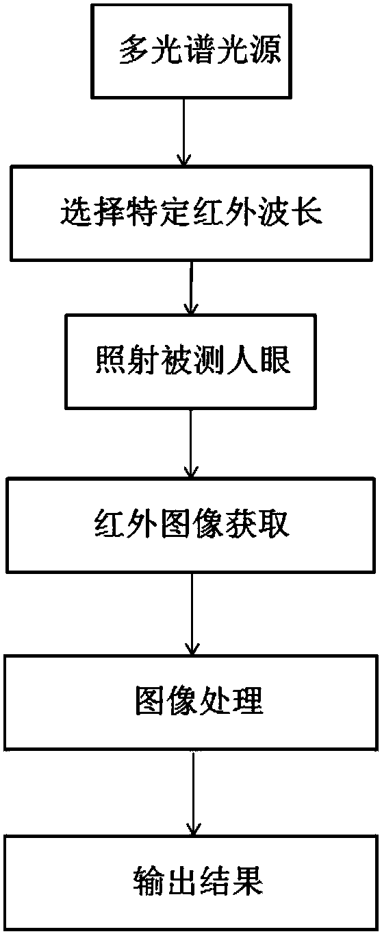
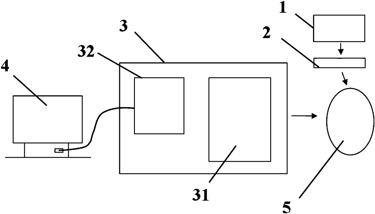
[0034] Such as figure 2 As shown, a multi-spectral meibomian gland detection and analysis device is provided with a multi-light source module 1 , a control module 2 , an image acquisition module 3 , and an image processing module 4 . The image acquisition module comprises a microscopic unit 32, a camera unit 31, and the objective lens of the microscopic unit 32 is aimed at the detected human eye, and one end of the reducing mirror is connected to the eyepiece of the microscopic unit 32, and the other end is connected to the camera unit 31, and the camera unit 31 just Images of the meibomian glands can be acquired directly. The image processing module 4 takes the images collected by the image collection module 3 as input, and uses a separation algorithm to enhance the contrast of the meibomian glands to make them clearly visible.

[0035] The specific operation process is as follows:...


PUM


Abstract

The invention provides a device for detecting a human eye meibomian gland model based on multispectrum and a method thereof, and relates to a medical optical detection device. The device for detecting a human eye meibomian gland model based on multispectrum is provided with a light source module, a control module, an image acquisition module, and an image processing module. The light source module is a multispectrum light source. Light emitted by the light source module passes a light filter group which is internally arranged in the light source module, the control module switches light filters, and light in specific central wavelength is output and irradiate human eyes. The image acquisition module comprises a camera unit and a microscopic unit, and the module is used to obtain meibomian gland images in the specific central wavelength and the images are input to the image processing module. The image processing module is provided with a separation algorithm, and analyzes and processes the meibomian gland images transmitted from the image acquisition module, to improve contrast of the meibomian gland images, and gland morphological parameters are calculated. The device distinguishes a meibomian gland and peripheral tissues, and the structure of the meibomian gland can be clearly observed.

Description

technical field [0001] The invention relates to a medical optical detection device, in particular to a device and method for detecting a human meibomian gland model based on multi-spectrum. Background technique [0002] The meibomian gland is a special differentiated sebaceous gland located in the tarsus of the eyelid, arranged vertically with the opening and the eyelid margin, and the lipid secretion discharged is evenly coated on the surface of the tear film through the blinking movement of the eyelid to form the tear film The outermost layer plays an important role in maintaining the health of the ocular surface: it can effectively delay the evaporation of the tear film water layer, reduce the surface tension of the tear film, enhance the stability of the tear film, prevent the skin of the eyelid margin from being soaked by tears, and provide smooth and smooth optics. Interface to reduce damage from blinking and act as a barrier to prevent contamination of the tear film w...

Claims


Application Information

Patent Timeline
Patent Type & Authority Patents(China)
IPC IPC(8): A61B5/00G06T7/00G06T5/00
CPCA61B5/0075A61B5/4222G06T5/00G06T7/0012G06T2207/10036G06T2207/30041
Inventor 陈延平郑天宇张洪桢杨伊璠刘孟珂孙步群
Owner XIAMEN UNIV
Why Eureka
Try Eureka