Endoscope probe and novel inherent fluorescence tumor diagnostic apparatus

An inherent fluorescence, tumor diagnosis technology, applied in the field of medical instruments, to achieve the effect of more convenient and more accurate detection

Inactive Publication Date: 2016-10-26
XUZHOU JINWEI MEDICAL ELECTRONICS EQUIP CO LTD
View PDF9 Cites 3 Cited by
  • Summary
  • Abstract
  • Description
  • Claims
  • Application Information

AI Technical Summary

Problems solved by technology

[0003] The original diagnostic method of inherent fluorescent tumor diagnostic instrument (patent number 02112880.4) is only for the screening of mucosal tumors on the surface of the cervix and vagina, while ignoring the possibility of malignant transformation of lesions such as the cervix and uterine cavity

Method used


Image

  • Endoscope probe and novel inherent fluorescence tumor diagnostic apparatus
  • Endoscope probe and novel inherent fluorescence tumor diagnostic apparatus
  • Endoscope probe and novel inherent fluorescence tumor diagnostic apparatus

Examples


Embodiment 1

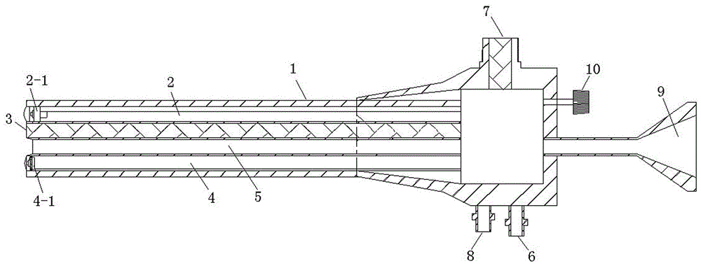
[0029] Such as figure 1 As shown, an endoscopic probe includes an optical fiber probe 1, and the optical fiber probe 1 is provided with an imaging channel 2, a fluorescence excitation light output channel 3, a white light output channel 4 and a biopsy channel 5, and a video camera is installed in the imaging channel 2 2-1, a white light source 4-1 is installed in the white light output channel 4; the rear end of the optical fiber probe 1 is provided with a camera signal output port 6, a fluorescence excitation light output port 7, a power port 8 and a biopsy channel entrance 9.

[0030] Camera 2-1 adopts a camera with no less than 795*596 pixels.

[0031] The rear end of the optical fiber probe 1 is also provided with a light adjustment knob 10, which can adjust the intensity of the fluorescence excitation light according to the detection environment or the clinical needs of the lesion to be detected.

Embodiment 2

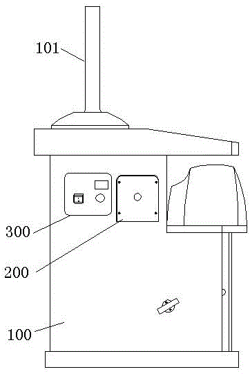
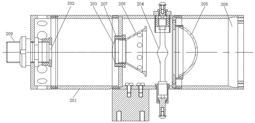
[0033] Such as figure 1 , figure 2 , image 3 As shown, a novel intrinsic fluorescence tumor diagnostic instrument includes a base 100, the upper surface of the base 100 is provided with a display 101, and the side of the base 100 is provided with an intrinsic fluorescence generator 200 and a fluorescence excitation light output terminal 300 , the inherent fluorescence generator 200 includes a housing 201, the inside of the housing 201 is sequentially arranged with a filter 202, a condenser 203, a mercury lamp 204, a reflector 205 and an axial fan 208, the filter 202, the condenser 203, the centerlines of the mercury lamp 204, the reflector 205 and the axial fan 208 are on the same straight line. A condenser cup 206 is arranged on the right side of the condenser lens 203 , and the condenser cup 206 is fixed together with the condenser lens 203 by a condenser lens fastener 207 . A fiber optic probe connector 209 is provided on the left side of the optical filter 202 . The ...


PUM


Abstract

The invention provides an endoscope probe. The endoscope probe comprises a light-guide fiber probe. A camera shooting channel, a fluorescence-excited light output channel, a white light output channel and a biopsy channel are arranged in the light-guide fiber probe. A camera is installed in the camera shooting channel, and a white light source is installed in the white light output channel. A camera signal output port, a fluorescence-excited light output interface, a power interface and a biopsy channel inlet are formed in the rear end of the light-guide fiber probe. Novel inherent fluorescence tumor diagnostic apparatus comprises the endoscope probe and a base. A displayer is arranged on the upper surface of the base, an inherent fluorescence generator and a fluorescence-excited light output end are arranged on the side face of the base, the fluorescence-excited light output interface is connected with the fluorescence-excited light output end, and the camera signal output port is connected with the displayer through a guide wire. According to the diagnostic apparatus, a former diagnosis method is abandoned, through perfect combination with a hysteroscope, precancerous lesions can be diagnosed earlier, and precancerous lesion tissue can be accurately found and taken out through the biopsy channel.

Description

technical field [0001] The invention relates to a medical instrument, in particular to an endoscopic probe and a novel intrinsic fluorescent tumor diagnostic instrument. Background technique [0002] A kind of material particles contained in normal human tissue and abnormal tissue are different. Different material particles produce fluorescence of different colors under fluorescence. Through different colors, it can be judged whether there is a lesion in the examined tissue, and early detection of tumors or precancerous lesions (such as CIN I CIN II). Guide biopsy positioning and find lesions when the mucosa is not ruptured. Intrinsic fluorescence is to macroscopically microscopic lesions from the perspective of molecular biology. [0003] The original diagnostic method of inherent fluorescent tumor diagnostic instrument (patent number 02112880.4) is only for the screening of cervical surface, vagina and other mucosal tumors, while ignoring the possibility of malignant tra...

Claims


Application Information

Patent Timeline
Patent Type & Authority Applications(China)
IPC IPC(8): A61B1/04A61B1/303A61B1/06A61B10/04
CPCA61B1/043A61B1/06A61B1/303A61B10/04A61B2576/02
Inventor 徐方张世潮庄金梅张玉松
Owner XUZHOU JINWEI MEDICAL ELECTRONICS EQUIP CO LTD
Why Eureka
Try Eureka