Gastrointestinal bleeding image detection method for capsule endoscopy

A technology for capsule endoscopy and gastrointestinal bleeding, which is applied in the field of image recognition, can solve the problems of not being able to identify old bleeding points well, and can not be well identified, so as to enhance adaptability, reduce workload, and improve recognition. rate effect

Active Publication Date: 2019-01-04
安翰科技(武汉)股份有限公司
View PDF5 Cites 2 Cited by
  • Summary
  • Abstract
  • Description
  • Claims
  • Application Information

AI Technical Summary

Problems solved by technology

This technology only uses red information (bleeding is generally red) when judging whether it is a bleeding image, and does not use any texture features
However, there are old bleeding spots in the bleeding image of the actual capsule endoscope. The color of this type of bleeding is not obvious red, but almost dark red, or even black, which cannot be well identified by this technology.
[0004] Prior art, "Research on Intelligent Recognition of Hemorrhage in Wireless Capsule Endoscope System and Endoscopic Image" (Pan Guobing, Ph.D. Dissertation), in which the method of hemorrhage recognition using color vector similarity coefficient and the method of hemorrhage recognition based on neural network are proposed , the two methods mentioned in the paper only use color information, and only using color information cannot identify old bleeding points very well

Method used


Image

  • Gastrointestinal bleeding image detection method for capsule endoscopy
  • Gastrointestinal bleeding image detection method for capsule endoscopy
  • Gastrointestinal bleeding image detection method for capsule endoscopy

Examples


Embodiment Construction

[0027] Below in conjunction with accompanying drawing and specific embodiment the present invention is described in further detail:

[0028] A kind of gastrointestinal bleeding image detection method for capsule endoscope of the present invention, it comprises the following steps:

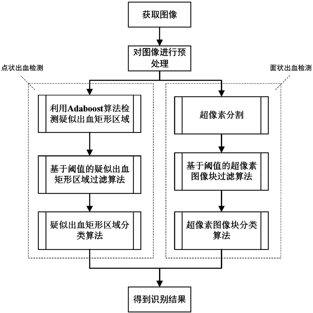
[0029] Step 1: After the patient swallows the capsule endoscope, the capsule endoscope collects images in the digestive tract and sends the images to the computer through wireless communication, and then the computer performs data preprocessing on the collected digestive tract images;

[0030] Step 2: Perform spotting detection and surface bleeding detection on the preprocessed digestive tract image to determine whether there is bleeding in the digestive tract, and mark the spot bleeding area and surface bleeding area;

[0031] The concrete steps of described spotting detection are:

[0032] Step 201: Using the Adaboost cascade algorithm based on Haar feature or LBP feature or Hog feature to train...


PUM


Abstract

The invention provides a digestive tract hemorrhage image detection method used for a capsule endoscope. The method comprises the following steps of 1: after a patient swallows the capsule endoscope, acquiring an image in the digestive tract by the capsule endoscope, transmitting the image to a computer through wireless communication, and performing data preprocessing on the digestive tract image by the computer; and 2: performing point-like hemorrhage detection and face-like hemorrhage detection on the preprocessed digestive tract image, determining whether hemorrhage exists in the digestive tract or not, and marking a point-like hemorrhage region and a face-like hemorrhage region. According to the method, the hemorrhage in the image can be effectively identified, so that the working efficiency of doctors is improved.

Description

technical field [0001] The invention relates to the technical field of image recognition, in particular to an image detection method for digestive tract bleeding used in a capsule endoscope. Background technique [0002] Gastrointestinal diseases, such as gastric cancer, intestinal cancer, acute and chronic gastritis, ulcer disease, etc., are mostly common diseases and frequently-occurring diseases, which pose a great threat to human health. Due to its high safety and high Reliability and other advantages, as an effective means of judging gastrointestinal diseases, it has been recognized by everyone and is widely used in gastrointestinal examination. Many diseases of the digestive tract are accompanied by bleeding. By combining computer technology and image recognition technology, the hemorrhage images in the image sequence can be accurately detected, which can assist doctors in diagnosing the digestive tract examination of the capsule endoscope system, improve the efficien...

Claims


Application Information

Patent Timeline
Patent Type & Authority Patents(China)
IPC IPC(8): G06T7/136G06T7/90
CPCG06T7/0012G06T2207/10068G06T2207/20081G06T2207/30028
Inventor 张行张皓袁文金王新宏段晓东肖国华
Owner 安翰科技(武汉)股份有限公司
Why Eureka
Try Eureka