

Biological image denoising algorithm based on statistical rule
An image and biological technology, which is applied in the field of optical microscopy and metrology microscopic imaging, can solve the problems of not meeting engineering needs and not being able to completely remove noise, and achieve the effect of improving the removal range and removal accuracy
- Summary
- Abstract
- Description
- Claims
- Application Information
AI Technical Summary
Problems solved by technology
Method used
Image

Examples
specific Embodiment approach 1
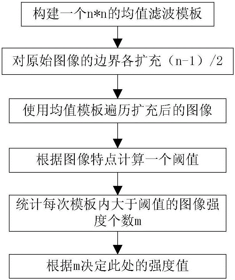
[0020] Specific Embodiment 1: The biological image denoising algorithm based on statistical laws described in this embodiment includes three steps of selecting a filter template, monitoring cell edges and noise, and image denoising processing. The specific process is:
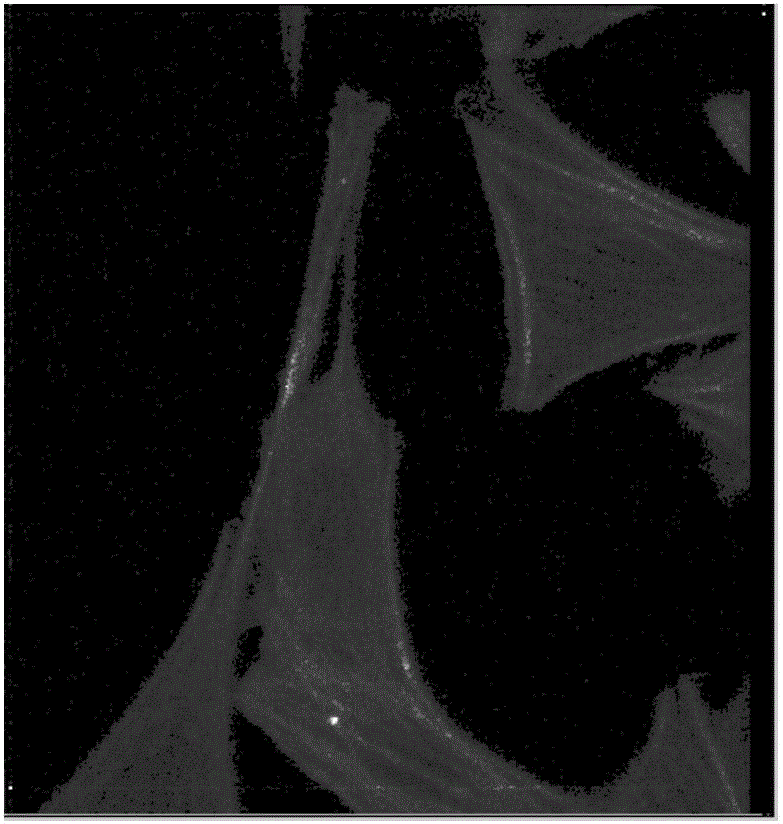
[0021] Step 1. Select a filter template: use a biological microscope to collect RGB images of biological cells, and select a filter template that can separate cells from isolated point noise by traversing the RGB images collected by the biological microscope. The selected filter templates are mean filter templates and Gaussian Filtering template, the RGB image is composed of a single cell and background area stray noise, or a cell group composed of multiple cells and background area stray noise;
[0022] Step 2, cell edge and noise monitoring: use the average filter template and Gaussian filter template to traverse the entire collected biological cell image, and determine the edge position of the biological cell...
specific Embodiment approach 2
[0026] Specific embodiment 2: This embodiment will further explain Embodiment 1. The specific process of using the mean filter template and the Gaussian filter template to traverse the entire biological cell image collected in step 2 to determine the position of the edge of the biological cell and the position of the isolated noise point is as follows: :
[0027] Use the mean filter template and the Gaussian filter template to traverse the entire collected biological cell image, use the Gaussian filter template in each region to obtain the filter threshold of the region, and then use the mean filter template to delete the larger than or If the number is smaller than the filtering threshold, it is determined whether the traversed area is an isolated point noise position or a cell edge or internal position according to the number.
specific Embodiment approach 3
[0028] Specific implementation mode three: this implementation mode further explains implementation mode two, assuming that the brightness value threshold S=48 obtained by the Gaussian filter template, when the brightness value inside the area traversed by the mean value filter template is greater than S=48, the number exceeds the mean value filter 1 / 2 of the template, the area traversed at this time is located at the edge or inside of the cell, and when the brightness value inside the area traversed by the mean filtering template is greater than S=48 and does not exceed 1 / 2 of the mean filtering template, this When traversing the region is located in the outlier noise position.
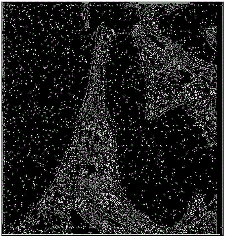
[0029] In this embodiment, the position of filtering or retaining the original image is selected according to the determined area division of the image, so as to achieve the effect of precise filtering. The image effect after filtering according to this method is as follows: Figure 4 shown.
[0030...
PUM
 Abstract
Description
Claims
Application Information