Optic disc segmentation method with combination of fundus image edge information and brightness information

A fundus image and edge information technology, which is applied in the field of medical image processing, can solve the problems of reducing the efficiency of the optic disc segmentation method, consuming a lot of time, and a large amount of calculation, and achieves fast positioning, improved efficiency, and high positioning accuracy.

Active Publication Date: 2017-03-22
TIANJIN UNIV
View PDF4 Cites 6 Cited by
  • Summary
  • Abstract
  • Description
  • Claims
  • Application Information

AI Technical Summary

Problems solved by technology

The method of optic disc segmentation by introducing vascular network information improves the robustness of the segmentation, but due to the complexity of the blood ve

Method used


Image

  • Optic disc segmentation method with combination of fundus image edge information and brightness information
  • Optic disc segmentation method with combination of fundus image edge information and brightness information
  • Optic disc segmentation method with combination of fundus image edge information and brightness information

Examples


Embodiment Construction

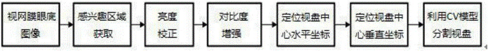
[0024] In order to make the purpose, technical solutions and advantages of the present invention clearer, the embodiments of the present invention will be further described in detail below in conjunction with the accompanying drawings:

[0025] (1): Preprocessing the fundus image

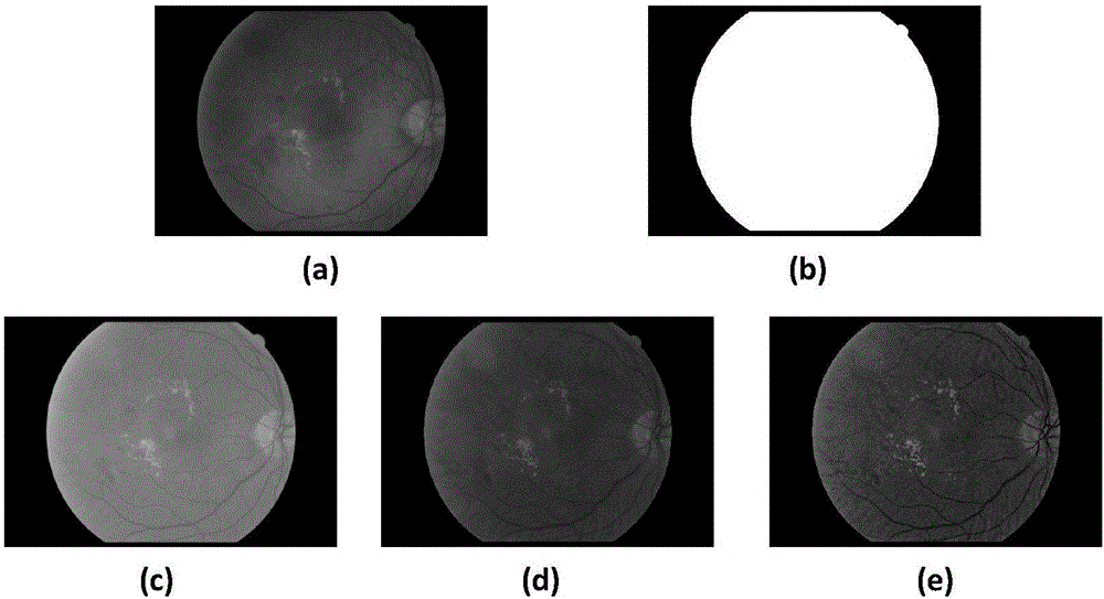
[0026] ①: First obtain the ROI of the fundus image. In this way, in the subsequent processing, the influence of pixels outside the ROI area can be effectively avoided, and the complexity of calculation can be reduced. Since the red component in the color fundus image is close to saturation and can best reflect the lighting conditions, the grayscale image I of the red channel component of the original image is selected r To process, take its maximum brightness value t max 5% of is used as a threshold for thresholding, and then a circular structuring element d with a radius of 3 3 The etching operation is performed to obtain the mask, and the result is as follows figure 2 (b), the formula is show...


PUM


Abstract

The invention relates to an optic disc segmentation method with the combination of fundus image edge information and brightness information. The method comprises a step of carrying out preprocessing on a fundus image, a step of using an Isotropic Sobel operator to extract the vertical edge and horizontal edge images of the fundus image with enhanced contrast, and positioning the horizontal coordinate of an optic disc according to an edge difference curve, a step of extracting a vascular intensiveness characteristic and an optic disc brightness characteristic, a step of defining the product of vascular intensiveness characteristic and the optic disc brightness characteristic as a comprehensive characteristic value, obtaining a comprehensive characteristic value curve about the vertical coordinate, wherein the coordinate corresponding to a maximum value in the curve is a optic disc vertical coordinate, and a step of cutting the optic disc by using a CV horizontal set model according to the positioned optic disc center coordinate. The method has high segmentation efficiency.

Description

technical field [0001] The invention belongs to the field of medical image processing, in particular to an optic disc segmentation method that integrates edge information and brightness information of fundus images. Background technique [0002] The automatic analysis of color retinal fundus images has been widely used in the auxiliary diagnosis of various ophthalmology-related systemic diseases such as diabetes, hypertension, kidney disease and neurological diseases. In a normal color fundus image, the macula, fovea, optic disc and vascular network are the four main features that can be observed in the fundus. The optic disc is in the shape of a light yellow or white disc, accounting for about 1% of the area of ​​interest in the entire fundus image. / 5 to 1 / 8, the blood vessels of the fundus diverge from the optic disc area and distribute in the whole fundus in a tree shape. The segmentation of the optic disc plays an extremely important role in the automatic analysis of f...

Claims


Application Information

Patent Timeline
IPC IPC(8): G06T7/12G06T7/13G06T5/30G06T5/00
CPCG06T5/007G06T5/30G06T2207/10024G06T2207/30041G06T2207/30101
Inventor 吕卫翟庆伟褚晶辉
Owner TIANJIN UNIV
Why Eureka
Try Eureka