X-ray phase imaging method based on imaging system features

An imaging system and imaging method technology, applied in the fields of biomedical engineering and medical imaging, can solve the problems of phase contrast image quality deterioration, phase contrast reduction, and the inability to guarantee the accuracy of phase information extraction results, etc.

Inactive Publication Date: 2017-04-26
TIANJIN UNIV
View PDF8 Cites 2 Cited by
  • Summary
  • Abstract
  • Description
  • Claims
  • Application Information

AI Technical Summary

Problems solved by technology

[0009] The gist of the present invention is to propose an X-ray phase imaging method based on the characteristics of the imaging system, so as to solve the key problems faced by the phase information extraction based on similar coaxial phase contrast imaging under the current engineering technology conditions: due to the current engineering conditions , X light source, non-ideality of the detector and system noise cause the quality of the phase contrast image to deteriorate, and the phase contrast is reduced. Based on the traditional phase extraction method, the accuracy of the phase information extraction result cannot be guaranteed.

Method used


Image

  • X-ray phase imaging method based on imaging system features
  • X-ray phase imaging method based on imaging system features
  • X-ray phase imaging method based on imaging system features

Examples


Embodiment Construction

[0024] The present invention will be described below in conjunction with the accompanying drawings and embodiments.

[0025] 1 Digital X-ray Imaging System

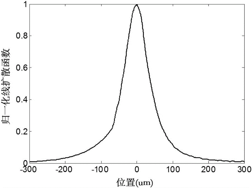
[0026] The experimental imaging system is Pixarray 100 small animal digital radiography system, manufactured by BIOPTICS Company of the United States. The detector of the system is a 1024×1024 CCD array with a pixel size of 50 μm×50 μm and 14 gray levels. The horizontal and vertical spatial resolutions are 20 pixels per millimeter. The focal spot size of the X-ray tube is 50 μm. In the experiment, the working voltage of the X-ray source is 33kVp, and the working current is 0.5mA. The imaging objects were constructed using polyethylene fibers with diameters of 100 μm, 50 μm, 20 μm and 10 μm. The experiment sets the distance from the X-ray source to the object as 100cm, and the corresponding distance from the object to the detector as 100cm. Under the above settings, the focal spot image formed by the light source on t...


PUM

PropertyMeasurementUnit
diameteraaaaaaaaaa

Abstract

The invention discloses an X-ray phase imaging method based on imaging system features. The X-ray phase imaging method comprises the following steps: setting X-ray in-line phase-contrast imaging parameters and exposure parameters of a digital radiography system, placing a tool edge device on an object plane to be imaged, acquiring a tool edge image, acquiring tool edge section curves at different positions in the image, and calculating a two-dimensional point function; placing an imaged object to image the object; calculating an initial value of a relaxation factor according to prior information about object construction and the noise condition of the obtained image; calculating phase information of the object; finely adjusting the relaxation factor according to the contrast of the obtained result until an ideal result is obtained.

Description

technical field [0001] The invention belongs to the fields of biomedical engineering and medical imaging, and relates to an X-ray phase imaging method based on the characteristics of an imaging system. Background technique [0002] Breast cancer is currently the number one "killer" of women. According to the latest statistics in 2015, the number of breast cancer patients and deaths worldwide increased to 1.7 million and 520,000 in 2012, accounting for 25% and 520,000 of all female cancer cases respectively. 15% of cancer deaths [2]. my country has a large population, and the prevalence of breast cancer has shown a clear upward trend in recent years. The diagnosis and treatment of breast cancer has become an increasingly serious social problem that needs to be solved urgently. Early diagnosis is the key to solving this social problem and improving the survival rate and life of patients. The key to quality. [0003] The current main method of routine breast examination is mol...

Claims


Application Information

Patent Timeline
Patent Type & Authority Applications(China)
IPC IPC(8): G01N23/04
CPCG01N23/04
Inventor 周仲兴高峰赵会娟张力新郭柏宽
Owner TIANJIN UNIV
Why Eureka
Try Eureka