
Application of fluorescent carbon dots in living cell nucleolus imaging or RNA labeling or display

A living cell nucleolus and fluorescent carbon dot technology, applied in the field of fluorescent carbon dots, achieves low cost, high practical value, and high imaging resolution

Inactive Publication Date: 2017-05-03
NINGBO INST OF MATERIALS TECH & ENG CHINESE ACADEMY OF SCI
View PDF7 Cites 12 Cited by
  • Summary
  • Abstract
  • Description
  • Claims
  • Application Information

AI Technical Summary

Problems solved by technology

So far, there has not been any report on the selective imaging effect of carbon dots on nucleoli

Method used


Image

  • Application of fluorescent carbon dots in living cell nucleolus imaging or RNA labeling or display
  • Application of fluorescent carbon dots in living cell nucleolus imaging or RNA labeling or display
  • Application of fluorescent carbon dots in living cell nucleolus imaging or RNA labeling or display

Examples


Embodiment 1

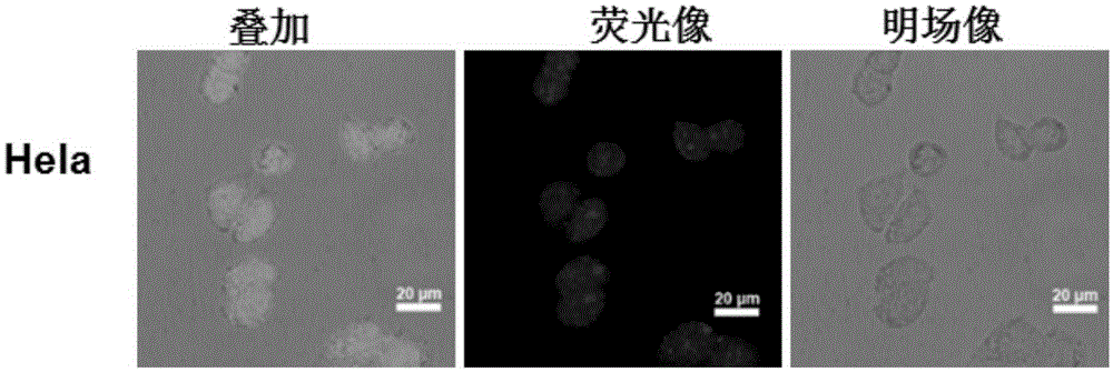
[0044] Example 1 Hela cells were stained with fluorescent carbon dots prepared by the method disclosed in the patent document with publication number CN104263366A. refer to figure 1 As shown, it can be seen that there are bright spots in the cells, and it is inferred that the bright spots are located in the nucleus.

Embodiment 2

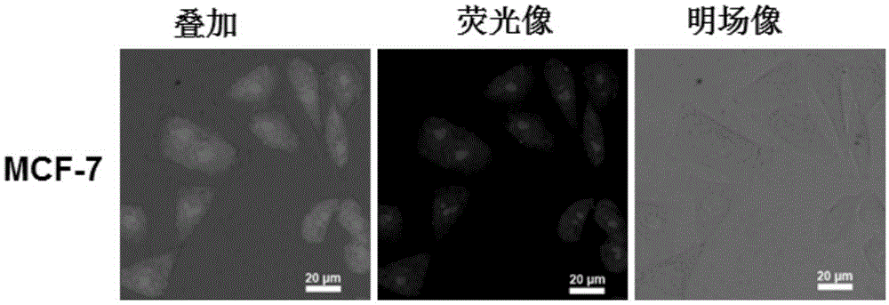
[0045] Example 2 MCF-7 cells were stained with fluorescent carbon dots prepared by the method disclosed in the patent document with publication number CN104263366A. refer to figure 2 As shown, it can be seen that there are bright spots in the cells, and it is inferred that the bright spots are located in the nucleus.

Embodiment 3

[0046] Example 3 In order to prove that the carbon dot staining area is located in the nucleus, fluorescent carbon dots prepared by the method disclosed in the patent document with publication number CN104263366A and Hoechst (nuclear specific dye) were used to co-stain Hela cells. refer to image 3 As shown, the blue Hoechst dye is located in the nucleus, and the superimposed picture shows that the red bright spot is located in the blue area, which can prove that the red bright spot is located in the nucleus.


PUM

PropertyMeasurementUnit
Particle sizeaaaaaaaaaa

Abstract

The invention discloses an application of fluorescent carbon dots in living cell nucleolus imaging or RNA labeling or display. The fluorescent carbon dots adopt red light emitting fluorescent carbon dots with an up-and-down conversion function, and particularly adopt fluorescent carbon dots disclosed in the CN104263366A. In some embodiments, the living cell nucleolus imaging can be accomplished by the use of the fluorescent carbon dots through cell culture, dye solution preparation, staining and imaging. With the help of the fluorescent carbon dots, the living cell nucleolus imaging or labeling or display of RNA in living cells can be realized, and the fluorescent carbon dots have the advantages of high selectivity on nucleolus imaging, high imaging resolution, simpliness, rapidness, low cost, low biological toxicity, and quite high practical value.

Description

technical field [0001] The invention relates to a fluorescent carbon dot, in particular to the application of a red light-emitting fluorescent carbon dot with up-down conversion function in cell nucleolus imaging or marking or displaying RNA in living cells, and belongs to the field of biotechnology. Background technique [0002] The nucleolus is an important structure in the nucleus. The nucleolus is the machine that produces ribosomes in the nucleus. The transcription of ribosomal RNA (rRNA) in all eukaryotes is completed in the nucleolus. The process is transcribed from rDNA to rRNA, and rRNA is then combined with proteins from the cytoplasm Combined, processed, transformed into ribosome precursors, and then exported to the cytoplasm. During the cell cycle, the nucleolus is a highly dynamic structure that exhibits periodic disappearance and reconstruction during mitosis. The nucleolus of eukaryotic cells has important functions as the site of rRNA synthesis, processing ...

Claims


Application Information

Patent Timeline
IPC IPC(8): G01N21/64
CPCG01N21/64
Inventor 林恒伟张凌孙山
Owner NINGBO INST OF MATERIALS TECH & ENG CHINESE ACADEMY OF SCI
Learn More