Image segmentation method and system

An image segmentation and medical image technology, which is applied in the field of medical image processing, can solve problems such as different sizes, variable texture shapes, and irregular edges on the shape, and achieve the effects of facilitating diagnosis and analysis, improving segmentation speed, and enhancing contrast

Inactive Publication Date: 2017-05-03
SHANGHAI UNITED IMAGING HEALTHCARE
View PDF4 Cites 26 Cited by
  • Summary
  • Abstract
  • Description
  • Claims
  • Application Information

AI Technical Summary

Problems solved by technology

[0005] In the prior art, the level set algorithm or multi-scale threshold method is used for nodule segmentation, but pulmonary nodules have different shapes, sizes, distribution locations, and are easily linked with other tissues, and their density is similar to that of some lung tissues. For example, pulmonary nodules have various forms such as solid nodules, mixed ground-glass nodules, and ground-glass nodules (GGN), and it is impossible to accurately identify pulmonary nodules, especially ground-glass nodules, simply by morphological methods. As a type of nodule with the highest possibility of malignancy, nodules have irregular edges in shape, and present fuzzy and thin shadows in CT images. The HU (Hounsfield Unit, Hounsfield Unit) value in CT is widely distributed and the texture morphology is variable. It is difficult to accurately identify pulmonary nodules by edge-based algorithms such as Level Set
In addition, the gray value of pulmonary nodules in the CT image does not obey the Gaussian distribution, and the multi-threshold method is easy to cause leakage and result in inaccurate segmentation results.

Method used


Image

  • Image segmentation method and system
  • Image segmentation method and system
  • Image segmentation method and system

Examples


Embodiment 1

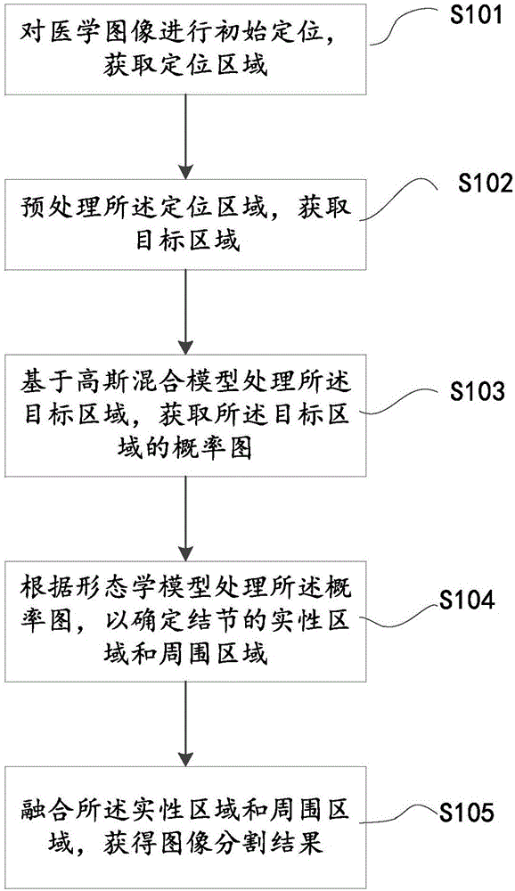
[0055] In order to solve the technical problem of effectively and accurately segmenting different types of lung nodules in the prior art, and to improve the accuracy of the user's diagnosis and analysis of the lesion, this embodiment provides an image segmentation method, such as figure 1 As shown in the schematic flow chart of the image segmentation method, the method includes the following steps:

[0056] Step S101 is performed: initial positioning is performed on the medical image, and the positioning area is acquired. The medical image medicine includes, but is not limited to, three-dimensional or two-dimensional images obtained through scanning and acquisition by various modal imaging systems, and can also be obtained through internal images such as a storage system image archiving and communication system (Picture Archiving and Communication System, PACS), etc. Or transfer to an external storage system. The modalities include, but are not limited to, one or more of magnetic...

Embodiment 2

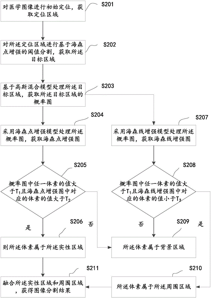
[0072] In order to make the above objectives, features and advantages more obvious and easy to understand, this embodiment provides a nodule segmentation method for three-dimensional CT images of the lungs to obtain different types of lung nodules, such as figure 2 As shown in the flowchart, the method includes the following steps:

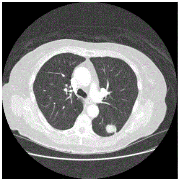
[0073] Step S201 is performed: initial positioning is performed on the medical image, and the positioning area is acquired. In this embodiment, the medical image is a lung medical image, and the medical image may be an original CT image obtained by scanning the human body by a computer tomography (CT) device, such as Figure 3a Shown. Or input the original CT image into a computer image processing device for processing, and obtain the required lung CT image based on methods such as threshold segmentation and clustering algorithms, such as Figure 3b Shown.

[0074] The initial positioning is used to obtain the positioning area, so as to reduce the cal...

Embodiment 3

[0092] In order to solve the above technical problems, an image segmentation system is provided in this embodiment. The image segmentation system may include one or more processing units, one or more storage units, one or more input units, and one or more output units. The units may be distributed or centralized, It can be local or remote.

[0093] In some embodiments, the input unit may respectively receive data sent from the imaging device, database, storage unit, or external device. The data here can be medical data. The medical data may be medical images. The medical image may include, but is not limited to, one or a combination of X-ray images, CT images, PET images, MRI images, ultrasound images, electrocardiograms, electroencephalograms, and the like. The medical image may be a two-dimensional (2D, two-dimensional) image or a three-dimensional (3D, three-dimensional) image. The format of the medical image may include, but is not limited to, Joint Photographic Experts G...


PUM


Abstract

The invention relates to an image segmentation method and system. The method comprises the following steps: initially positioning medical images to obtain a positioning area; preprocessing the positioning area to obtain a target area, wherein the target area contains a nodular area and a background area, and the nodular area consists of a solid area and a surrounding area which surrounds the solid area; processing the target area in combination with a Gaussian mixture model to obtain a probability graph of the target area; processing the probability graph according to a morphological model to determine the solid area and the surrounding area of a nodule; and fusing the solid area and the surrounding area to obtain an image segmentation result. According to the method and system provided by the invention, different types of nodules can be accurately segmented, and the follow-up diagnostic analysis can be effectively improved.

Description

[0001] 【Technical Field】 [0002] The present invention relates to the field of medical image processing, in particular to an image segmentation method and system. [0003] 【Background technique】 [0004] Lung cancer is the cancer with the highest mortality rate in the world. Although the level of medical diagnosis and treatment continues to improve, the five-year survival rate of lung cancer is only about 15%. Early detection and early treatment are the main ways to improve the cure rate of lung cancer. Lung nodules are an early manifestation of lung cancer. The accurate segmentation results of lung nodules can effectively reflect the pathological and morphological characteristics of the nodules, thereby helping users to diagnose and analyze the lesions. The key technology to realize the automatic analysis and recognition of lung nodules is the research and application of a series of image processing, analysis and understanding algorithms such as lung nodule segmentation, detectio...

Claims


Application Information

Patent Timeline
Patent Type & Authority Applications(China)
IPC IPC(8): G06T7/00G06T7/11G06T7/136G06T7/194
CPCG06T7/0012G06T2207/10081G06T2207/20101G06T2207/30064
Inventor 王季勇
Owner SHANGHAI UNITED IMAGING HEALTHCARE
Why Eureka
Try Eureka