
Biocompatible built-in stent material

A technology of biocompatibility and prosthetic materials, applied in drug delivery, surgery, microcapsules, etc., can solve the problems of loss of EC function, vascular stenosis, and poor clinical effect of endothelial inoculation, so as to avoid restenosis and inhibit restenosis Effect

Active Publication Date: 2017-05-31
CHINABRIDGE (SHENZHEN) MEDICAL TECH CO LTD
View PDF14 Cites 0 Cited by
  • Summary
  • Abstract
  • Description
  • Claims
  • Application Information

AI Technical Summary

Problems solved by technology

Synthetic grafts have also been inoculated with endothelial cells, but the clinical outcome of endothelial inoculation is generally poor, most likely due to nonadhesive properties of the cells to the graft and / or loss of EC function due to ex vivo manipulation
CN105327399 provides a method for constructing artificial blood vessels, which introduces prokaryotic system expression vectors with hydrophilic and negatively charged polypeptide genes and cell adhesion-promoting polypeptide genes on the surface (Journal of DonghuaUniversity (English Edition), 2012, 29:26 -29; Bio-Medical Materials and Engineering, 2014, 24:2057–2064), which improves surface hydrophilicity and negative charge, increases endothelialization potential, and is beneficial to tissue healing and anticoagulation, but this technology is suitable for constructing artificial blood vessels, The materials used include polyester. Obviously, for the field of vascular stents that need to be degraded after vascular stenosis treatment, this material has natural non-biocompatibility and cannot provide the mechanical support properties required by vascular stents. At the same time, as mentioned above As mentioned above, if endothelial cell proliferation cannot maintain homeostasis, it will lead to the formation of vascular restenosis

Method used


Image

  • Biocompatible built-in stent material
  • Biocompatible built-in stent material
  • Biocompatible built-in stent material

Examples

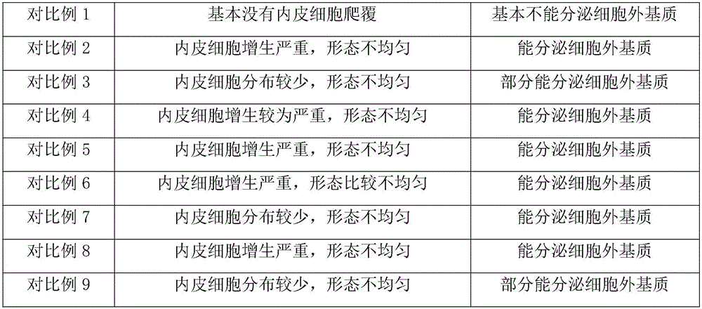
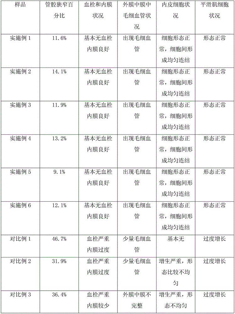

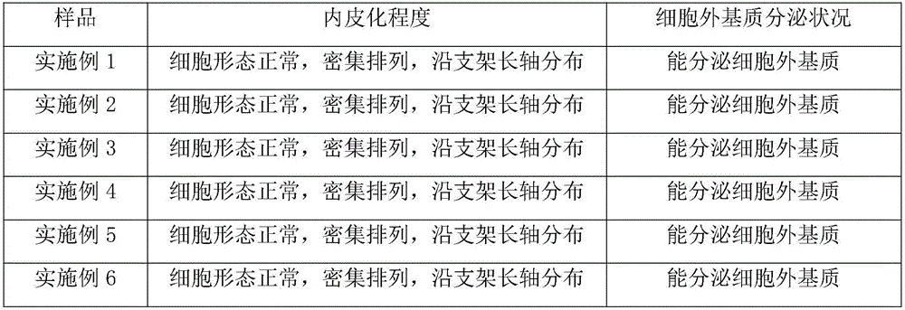
Embodiment 1

[0051] Preparation of prostheses (preferably endovascular stents) with microcavities and short peptide coatings

[0052] Step 1: Obtain the base material of the prosthesis, which can degrade the pure iron tubular material, and make it have holes connecting the inner and outer surfaces on the surface;

[0053] Step 2: Using laser engraving technology, a microcavity is obtained in the thickness direction of the above-mentioned hole that runs through the prosthesis; the microcavity has a hemispherical outline, the diameter of which is 30 μm, and the depth of the bottom is the diameter of the hole 1 / 3 of , and the total number of the microcavities is 30;

[0054] Step 3: Using conventional techniques, obtain a composition in the form of microcapsules, the composition comprising gelatin as a "shell", and histidine chelated iron as a "core", wherein the histidine chelated iron is in the composition The weight content of the gelatin is 0.5%, and the degradation cycle of the gelatin ...

Embodiment 2

[0059] Preparation of prostheses (preferably endovascular stents) with microcavities and short peptide coatings

[0060] The preparation process is the same as that in Example 1, except that: the matrix material used in step 1 is degradable polycaprolactone; in step 2, the diameter of the microcavity is 50 μm, and the depth dimension of the bottom is 1 / 4 of the diameter of the hole, The total number of microcavities is 40; in step 3, the shell is chitosan, the degradation period is about 1 week, the core is cysteine ​​chelated iron, and the weight content of the core in the composition is 0.8%; The loading in step 4 was 8 μg.

Embodiment 3

[0062] Preparation of prostheses (preferably endovascular stents) with microcavities and short peptide coatings

[0063] The preparation process is the same as in Example 1, except that: the matrix material used in step 1 is degradable pure magnesium; the diameter of the microcavity in step 2 is 100 μm, the depth dimension of the bottom is 1 / 5 of the diameter of the hole, and the microcavity is 1 / 5 of the diameter of the hole. The total number is 60; in step 3, the shell is collagen, the degradation period is about 2 weeks, the core is cysteine ​​chelated iron, and the weight content of the core in the composition is 1.5%; in step 4, the load is The amount is 10 μg; the sequences of the two short peptides in step 5 are the aforementioned sequence 3 and sequence 4, respectively.


PUM

PropertyMeasurementUnit
thicknessaaaaaaaaaa
diameteraaaaaaaaaa
diameteraaaaaaaaaa

Abstract

The invention relates to a biocompatible endoprosthesis material, which comprises a matrix, a microcavity, a composition and short-peptide layers, wherein the material of the matrix is biodegradable in vivo tissues, the matrix is in a tubular shape and is provided with an inner tubular surface and an outer tubular surface, the inner surface forms a channel for a tissue fluid such as blood to pass through, and a structure for communicating the inner surface and the outer surface is arranged on the surface; the microcavity is obtained when a communicating structure is located in a cross-section direction, perpendicular to an axial direction, of a matrix body; the microcavity is located in a thickness direction of a hole; a plane of an opening of the microcavity is basically perpendicular to an inner surface and an outer surface of a tubular-shaped stent; the composition is loaded in the microcavity and can be degraded to release Fe<3+>; the short-peptide layers are located on the inner surface and the outer surface of the matrix material; short peptides can be self-assembled to form hydrogel; the hydrogel formed after the short peptides are self-assembled can be specifically favorable to climbing and covering of endothelial cells and regeneration of endodermis; a prosthesis can provide required biocompatibility, has balanced climbing and covering of an EC layer while keeping the support force of the stent, inhibits restenosis, and recovers tissues, especially the self performance of a blood vessel at the right moment.

Description

Background technique [0001] The invention belongs to the field of medical materials, and relates to a prosthesis material, in particular, a scaffold material with biocompatibility, more suitable for intravascular scaffold material. [0002] The body includes various passages, such as arteries, other blood vessels, and other body lumens. Sometimes these channels become blocked or weakened. For example, the channel can be blocked by a tumor, narrowed by plaque, or weakened by an aneurysm. When these conditions occur, the channel can be reopened or augmented, or even replaced, using a medical endoprosthesis. An endoprosthesis is typically a tubular member placed in a lumen in the body. Examples of endoprostheses include stents, covered prostheses, graft stents, and vessel closure pins. [0003] Stents (stents, stents), venous filters, expandable frames and similar implantable medical devices, hereinafter collectively referred to as stents, are radially expandable endoprosthes...

Claims


Application Information

Patent Timeline
Patent Type & Authority Applications(China)
IPC IPC(8): A61L31/14A61L31/16A61L31/04
CPCA61L31/047A61L31/145A61L31/148A61L31/16A61L2300/102A61L2300/416A61L2300/604A61L2300/622C08L89/00
Inventor 石佳明
Owner CHINABRIDGE (SHENZHEN) MEDICAL TECH CO LTD
Learn More