Method for detecting DNA variation level by using polymolecular tags

A label detection and multi-molecular technology, applied in the field of DNA variation detection, can solve the problems of indistinguishability, data waste, and high cost, and achieve the effect of reducing cost, improving accuracy, and lowering the threshold

Inactive Publication Date: 2017-06-27
苏州首度基因科技有限责任公司 +1
View PDF3 Cites 10 Cited by
  • Summary
  • Abstract
  • Description
  • Claims
  • Application Information

AI Technical Summary

Problems solved by technology

[0006] However, the current UID technology cannot distinguish whether the amplified sequence from the same double-stranded DNA template contains both strands of the double strand; and the prior art uses a fully synthetic method to prepare barcode, which is very e

Method used


Image

  • Method for detecting DNA variation level by using polymolecular tags
  • Method for detecting DNA variation level by using polymolecular tags
  • Method for detecting DNA variation level by using polymolecular tags

Examples


Embodiment 1

[0039] 1. Synthesis and annealing of the adapter

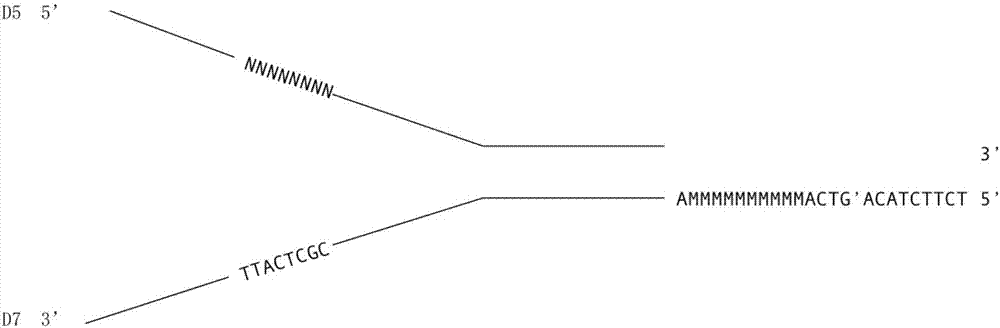
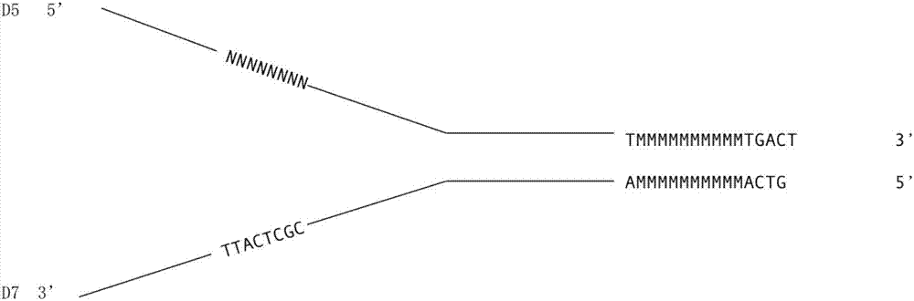
[0040] 1) Synthesize the following sequences in Table 1, wherein NNNNNNNNN in J-AdD5-8N represents the first DNA molecule tag N with a length of 8bp 1 , in J-AdD703-10M MMMMMMMMMM Represents the second DNA molecule label N with a length of 10bp 2 , where the third DNA molecule labels N 3 It is a pre-synthesized short fragment, which is the bold base sequence CGCTCATT in this sequence.

[0041] Table 1

[0042]

[0043] 2) Centrifuge to collect the Adapter powder at the bottom of the tube.

[0044] 3) Open the Adapter tube cap carefully, and be careful not to let the powder float out. Add the corresponding volume of OAB dissolved powder to J-AdD5-8N and J-AdD703-10M respectively, so that the final concentration is 100 μM, and store the stock solution at -20°C for future use. OAB Buffer (oligomeric annealing buffer), the composition is shown in Table 2:

[0045] Table 2

[0046]

[0047] 4) Take 25 μL of J-AdD5-8N...

Embodiment 2

[0076] In the synthesis and annealing reaction of the adapter (adapter) in the first step, the following sequences in Table 7 were synthesized, wherein CGCTCATT in J-AdD5 represents the first DNA molecule tag N with a length of 8bp 1 , in J-AdD703-8N-10N NNN NNN NNNN Represents the second DNA molecule label N with a length of 10bp 2 , NNNNNNNN represents the third DNA molecular label N with a length of 8bp 3 , the synthetic DNA sequence is as follows:

[0077]

[0078] All the other steps are the same as in Example 1.

Embodiment 3

[0080] In the synthesis and annealing reaction of the adapter (adapter) in the first step, the following sequences in Table 8 were synthesized, wherein NNNNNN in J-AdD5-6N represents the first DNA molecule tag N with a length of 6bp 1 , NNNNNN in J-AdD703-6N represents the second DNA molecule label N with a length of 6bp 2 , where the third DNA molecule labels N 3 It is a pre-synthesized short fragment, which is the bold base sequence CGCTCATT in this sequence.

[0081] Table 8

[0082]

[0083] All the following steps are the same as in Example 1. The first DNA molecular label N in each of the above embodiments 1 Sequence, second DNA molecule tag N 2 Sequence and third DNA molecule label N 3 The bases in the sequence are randomly selected from A, T, C and G.


PUM


Abstract

The invention relates to a method for detecting a DNA variation level by using polymolecular tags. The method comprises the following steps: inserting a first DNA tag N1 into D5 so as to obtain a first single stranded DNA molecule, wherein the sequence length of the N1 is 2-12bp; inserting a second DNA tag N2 and a third DNA tag N3 into D7 so as to obtain a second single stranded DNA molecule, wherein sequence lengths of the N2 and N3 are respectively 2-12bp and 2-20bp; mixing the two single stranded DNA molecules, annealing, carrying out a DNA polymerization reaction, and performing digestion by using restriction enzymes so as to obtain a DNA linker sequence containing a first sticky end; breaking to-be-detected DNA into short DNA fragments, and adding a second sticky end in reverse complement with the first sticky end at two ends of the DNA fragments; and connecting the DNA linker sequence and the short DNA fragments, performing PCR (Polymerase Chain Reaction) and sequencing, contrasting with human reference genome so as to judge the variation level of the to-be-detected DNA.

Description

technical field [0001] The invention relates to the field of DNA variation detection, in particular to a method for detecting DNA variation level with multi-molecular labels. Background technique [0002] Compared with traditional detection methods, liquid biopsy technology is a hot technology in the field of tumor precision medicine because of its advantages of less side effects, simple operation, and repeatable sampling. Its main detection objects include: circulating tumor cells (CTCs), circulating Tumor DNA (ctDNA) and tumor exosomes can prompt information such as tumor development and drug resistance, and guide individualized drug treatment. [0003] ctDNA is a kind of free extracellular single-stranded or double-stranded DNA existing in plasma, serum, cerebrospinal fluid and other body fluids, mainly from necrotic or apoptotic tumor cells, exosomes secreted by tumor cells, and circulating tumor cells. The size of ctDNA fragments is usually 160-180bp, with a half-life ...

Claims


Application Information

Patent Timeline
IPC IPC(8): C12Q1/68
CPCC12Q1/6869C12Q2525/191C12Q2531/113C12Q2565/102
Inventor 王海龙徐健唐元华
Owner 苏州首度基因科技有限责任公司
Why Eureka
Try Eureka