PET imaging system and imaging method thereof

An imaging method and image technology, applied in the medical field, can solve the problems of poor user experience, long scanning and image reconstruction time, etc., and achieve the effects of short scanning time, shortening image reconstruction time, and improving efficiency

Pending Publication Date: 2017-07-21
SHANGHAI UNITED IMAGING HEALTHCARE
View PDF5 Cites 9 Cited by
  • Summary
  • Abstract
  • Description
  • Claims
  • Application Information

AI Technical Summary

Problems solved by technology

This method not only takes a long time for scanning and image reconstruction, but also has poor user experience, and for positions without respiratory movement such as legs, the time spent on scanning and the obtained PET data are mostly meaningless clinically.

Method used


Image

  • PET imaging system and imaging method thereof
  • PET imaging system and imaging method thereof
  • PET imaging system and imaging method thereof

Examples


Embodiment 1

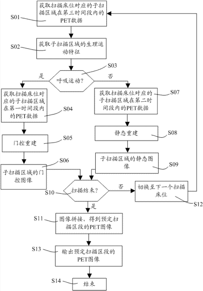
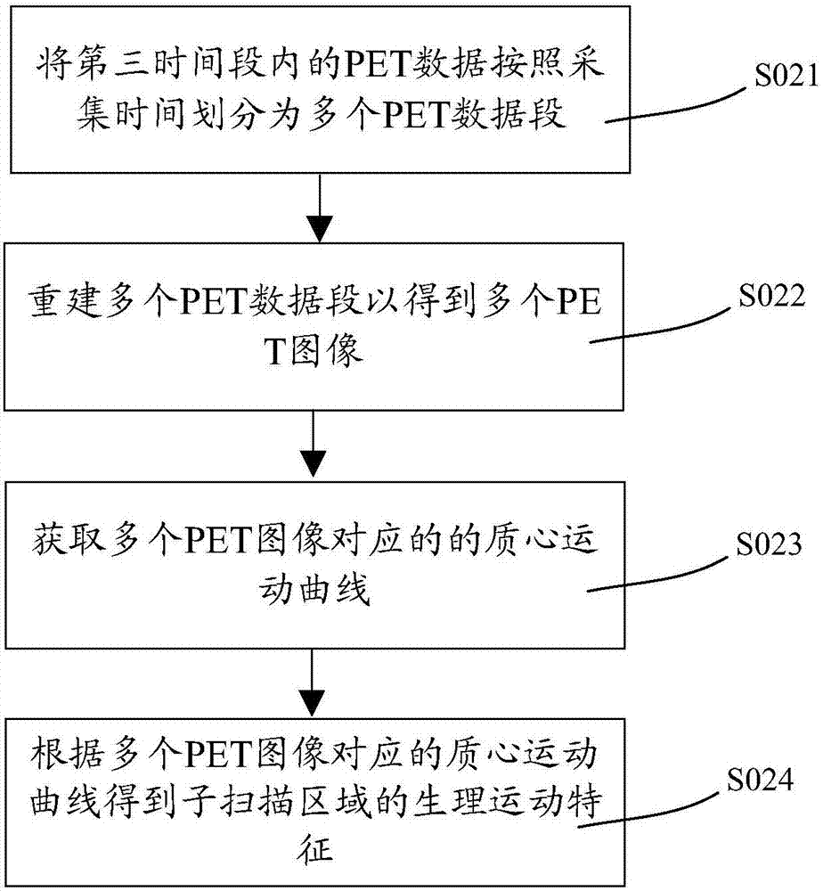
[0056] figure 1 It is a flow chart of the PET imaging method provided by Embodiment 1 of the present invention, such as figure 1 As shown, the PET imaging method specifically includes the following steps.

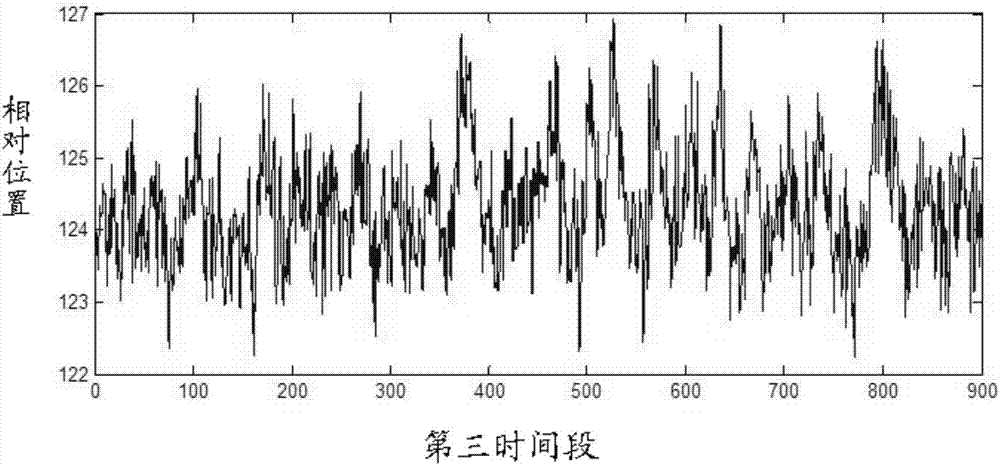
[0057] Step S01 is: acquiring PET data of a sub-scan area corresponding to a scan bed within a third time period.

[0058] It should be understood that each sub-scanning area corresponds to a scanning bed, for example, a patient's head corresponds to a scanning bed, the patient's chest and abdomen correspond to another scanning bed, and the patient's lower limbs correspond to the rest of the scanning bed. The corresponding scanning beds constitute double beds. Therefore, the sub-scanning area can be adjusted by changing the scanning bed position. One scanning bed corresponds to one PET scan. After the scanning of one scanning bed is completed, the patient is placed on the examination bed to move to the next scanning bed for PET scanning, that is, the sub-scanning area co...

Embodiment 2

[0103] The principle of the PET imaging method provided in this embodiment is basically the same as that in Embodiment 1, and only the differences will be described below. In this embodiment, a user ID is set for at least one scanning bed, so as to determine whether there is physiological movement in the sub-scanning area corresponding to the scanning bed by reading the user ID of the at least one scanning bed. For example, the same user ID is set for the scanning bed corresponding to the patient's head and the scanning bed corresponding to the patient's lower limbs, and another user ID is set for the scanning bed corresponding to the patient's chest and abdomen, and the two user IDs are different.

[0104] Concrete combination Figure 5 In other words, the PET imaging method of this embodiment replaces the steps S01, S02 and S03 in the first embodiment with steps S21 and S22, and the rest of the steps are the same as those in the first embodiment.

[0105] Wherein, step S21 ...

Embodiment 3

[0109] The principle of the PET imaging method provided in this embodiment is basically the same as that in Embodiment 1, and only the differences will be described below.

[0110] combine Image 6 In other words, the PET imaging method of this embodiment replaces the steps S01, S02 and S03 in the first embodiment with steps S31 and S32, and the rest of the steps are the same as those in the first embodiment.

[0111] Wherein, step S31 is: acquiring a CT positioning image of the sub-scanning area corresponding to the scanning bed.

[0112] Step S32 is: according to the CT positioning image, identify whether there is respiratory movement in the sub-scanning area. Here, if the CT positioning image presents a sub-scan area with physiological movement, then step S04 is performed, otherwise, step S07 is performed.

[0113] In this embodiment, before judging whether there is respiratory movement in the sub-scanning area, the CT positioning image (Topogram, also called Topo image) ...


PUM


Abstract

The invention relates to a PET imaging system and an imaging method thereof. The PET imaging method comprises the following steps: dividing a predetermined scanning section into one or more sub-scanning areas, wherein each sub-scanning area corresponds to a scanning bed; prejudging whether or not there is physiological movement in the corresponding sub-scanning area for each scanning bed; if yes, obtaining PET data of the sub-scanning area corresponding to the scanning bed in a first time period through a first data acquisition mode and obtaining the gated image of the sub-scanning area at least based on the PET data in the first time period; if not, obtaining the PET data of the sub-scanning area corresponding to the scanning bed in a second time period through a second data acquisition mode and obtaining the static image of the sub-scanning area according to the PET data in the second time period. The PET imaging system comprises a dividing unit, a judging unit, a scanning unit and a reconstruction unit, and is used for obtaining the static or gated image of the sub-scanning area through the imaging method.

Description

technical field [0001] The invention relates to the field of medical technology, in particular to a PET imaging system and an imaging method thereof. Background technique [0002] PET (Positron Emission Tomography: Positron Emission Tomography) imaging technology uses drugs such as labeled glucose, amino acid, thymine, receptor ligands or blood flow imaging agents as tracers, in the form of anatomical images, from The metabolism, function, blood flow, cell proliferation or receptor distribution of the body and lesion tissue cells on the molecular level can provide more physiological and pathological diagnostic information for the clinic, and have excellent performance in helping the early diagnosis of cancer. [0003] Among them, data acquisition is an important part of the PET system. There are many working methods for PET data acquisition, but in general, PET data acquisition includes three scanning methods, namely transmission scanning, gated scanning and static scanning....

Claims


Application Information

Patent Timeline
Patent Type & Authority Applications(China)
IPC IPC(8): A61B6/03
CPCA61B6/032A61B6/037A61B6/5235A61B6/527A61B6/54A61B6/4417A61B6/461A61B6/5205A61B6/5264A61B6/545A61B6/563A61B6/541A61B6/0487G06T11/005G06T2207/10104G06T2211/412
Inventor 孙友军冯涛
Owner SHANGHAI UNITED IMAGING HEALTHCARE
Why Eureka
Try Eureka