Ultrasonic contrast imaging method and system

A technology of contrast-enhanced ultrasound and imaging methods, applied in ultrasound/sonic/infrasonic diagnosis, sonic diagnosis, infrasonic diagnosis, etc., can solve problems such as low success rate and inaccurate registration results, reduce impact, increase clinical diagnosis confidence, The effect of improving the registration success rate

Active Publication Date: 2017-07-21
SHENZHEN MINDRAY BIO MEDICAL ELECTRONICS CO LTD +1
View PDF6 Cites 19 Cited by
  • Summary
  • Abstract
  • Description
  • Claims
  • Application Information

AI Technical Summary

Problems solved by technology

[0004] Based on this, it is necessary to provide a new contrast-enhanced ultrasound imaging method and system for the existing problem

Method used


Image

  • Ultrasonic contrast imaging method and system
  • Ultrasonic contrast imaging method and system
  • Ultrasonic contrast imaging method and system

Examples


Embodiment Construction

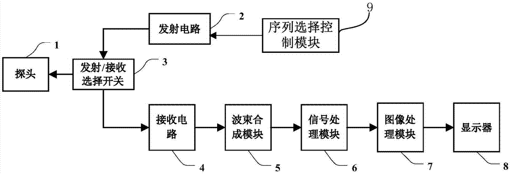
[0031] Such as figure 1 As shown, the device for ultrasonic imaging of the target area according to the embodiment of the present invention includes: a probe 1, a transmitting circuit 2, a transmitting / receiving selection switch 3, a receiving circuit 4, a beam forming module 5, a signal processing module 6, and an image processing module 7 and display 8.

[0032] The transmitting circuit 2 transmits the delayed-focused ultrasonic pulse with a certain amplitude and polarity to the probe 1 through the transmitting / receiving selection switch 3 . Probe 1 is excited by ultrasonic pulses, and emits ultrasonic waves to the target area (not shown in the figure) of the body tissue under test, and receives the ultrasonic echoes with tissue information reflected from the target area after a certain delay, and sends the The ultrasound echoes are converted back into electrical signals. The receiving circuit receives the electrical signal converted and generated by the probe 1 to obtain ...


PUM


Abstract

The invention relates to an ultrasonic contrast imaging method and system. The system comprises a probe 1, a transmission circuit 2, a reception circuit 4 and a signal processing module, wherein the transmission circuit is used for respectively transmitting a first pulse sequence and a second pulse sequence to a target area through the probe; the reception circuit is used for respectively receiving ultrasonic echoes of the first pulse sequence and the second pulse sequence through the probe so as to obtain a first group of ultrasonic echo signals and a second group of ultrasonic echo signals; and the signal processing module is used for extracting echo signal components according to the first group of ultrasonic echo signals and the second group of ultrasonic echo signals. The invention furthermore discloses a new ultrasonic contrast image transmission control method, so that the registering success rate of a software analysis tool is improved.

Description

technical field [0001] The invention relates to the field of medical ultrasound imaging, in particular to a method and system for performing ultrasound contrast imaging on a target area. Background technique [0002] Ultrasound contrast agent microbubbles can enhance the intensity of reflected echoes. Its diameter is as small as the size of blood cells, and it can spread to various organs of the human body along with the blood of the human body. Microbubble movement has significant nonlinear characteristics. With the maturity of contrast-enhanced ultrasound imaging technology based on nonlinear characteristics, contrast-enhanced ultrasound imaging has been widely used in the clinical identification, diagnosis and treatment of benign and malignant tumors. The perfusion performance of ultrasound-enhanced microbubbles over time has formed a unified clinical standard in the differentiation of benign and malignant liver tumors. In recent years, with the development of interventi...

Claims


Application Information

Patent Timeline
IPC IPC(8): G06F19/00A61B8/00
Inventor 桑茂栋丛龙飞
Owner SHENZHEN MINDRAY BIO MEDICAL ELECTRONICS CO LTD
Why Eureka
Try Eureka