Multi-path, multi-magnification, non-confocal fluorescence emission endoscopy apparatus and methods

a fluorescence emission endoscopy and multi-magnification technology, applied in the field of multi-photon fluorescence and/or nonlinear harmonic emission endoscopy apparatus and methods, can solve the problems of inconvenient use of high-energy light, inability to provide a compact endoscope, and inability to adapt to the architecture of switchable optical systems

Active Publication Date: 2010-10-14
CORNELL UNIVERSITY
View PDF44 Cites 101 Cited by
  • Summary
  • Abstract
  • Description
  • Claims
  • Application Information

AI Technical Summary

Benefits of technology

[0024]The embodiments described herein take advantage of the fact that the fluorescence emission excitation field is typically in the near IR (˜700 nm<λ≦1.3μ) spectrum; thus the excitation wavelength can be easily separated from the substantially shorter fluorescence emission wavelengths and the visible light (−400 nm≦λ≦700 nm) for viewing at low magnification. Furthermore, since the 3D resolution of multiphoton imaging results entirely from the scanned laser excitation, the fluorescence emission collection optical system requires only the capability to provide efficient emitted signal collection, facilitating the use of a simple, large aperture lens rather than a highly corrected objective lens. The non-confocal design of the high-magnification optical system provides added advantage for imaging deep in tissue because the problem of image obstruction from a concentric fiber scanner is reduced, as is the concern that the excitation fiber will not provide sufficiently efficient signal collection and / or transmission.
[0026]In various exemplary applications, the apparatus and method embodiments described herein can be used to provide in vivo, in situ microscopic imaging of the infrastructure and bio-chemistry of tissue as a shortcut to diagnostic information that ordinarily would be later acquired by a pathologist's hematoxylin and eosin (H&E) stained absorption microscopy of fixed thin slices of tissue obtained by biopsy. The embodied apparatus and methods of the present invention may be utilized during surgery or during precursor diagnostics to delineate boundaries of malignant tumors or to recognize particular disease or damaged states, which will inform their prompt treatment, and for monitoring past treatment results. The embodied apparatus and methods may be used to identify and recognize organs and anatomical structures to be protected from injury during surgery, such as nerve bundles, microtubule bundles, and other structures. The embodied apparatus and methods will allow accurate spatial target discrimination, and permit non-imaging quantification of fluorescence emission from target volumes.

Problems solved by technology

The use of high energy light could easily damage living tissue in the entire region of exposure.
Although multi-optical systems are routinely provided in microscopy apparatus, separate, switchable optical systems do not provide a suitable architecture for a compact endoscope.
The inventors have also recognized that conventional confocal-based endoscopy imaging apparatus and methods can be disadvantageous due, for example, to image obstruction from scanner apparatus and inefficient signal collection and / or transmission via a concentric excitation fiber waveguide.

Method used


Image

  • Multi-path, multi-magnification, non-confocal fluorescence emission endoscopy apparatus and methods
  • Multi-path, multi-magnification, non-confocal fluorescence emission endoscopy apparatus and methods
  • Multi-path, multi-magnification, non-confocal fluorescence emission endoscopy apparatus and methods

Examples


Embodiment Construction

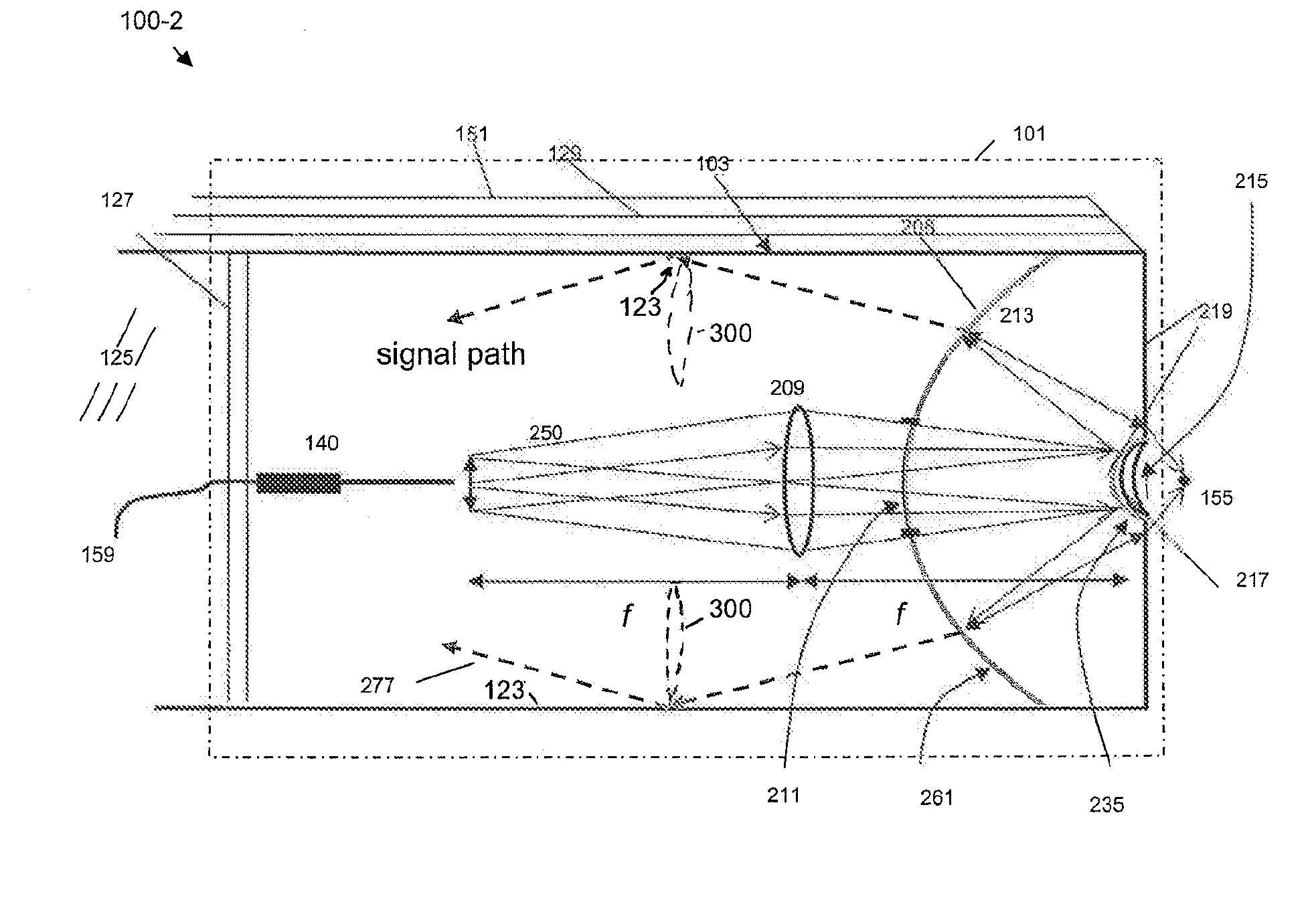
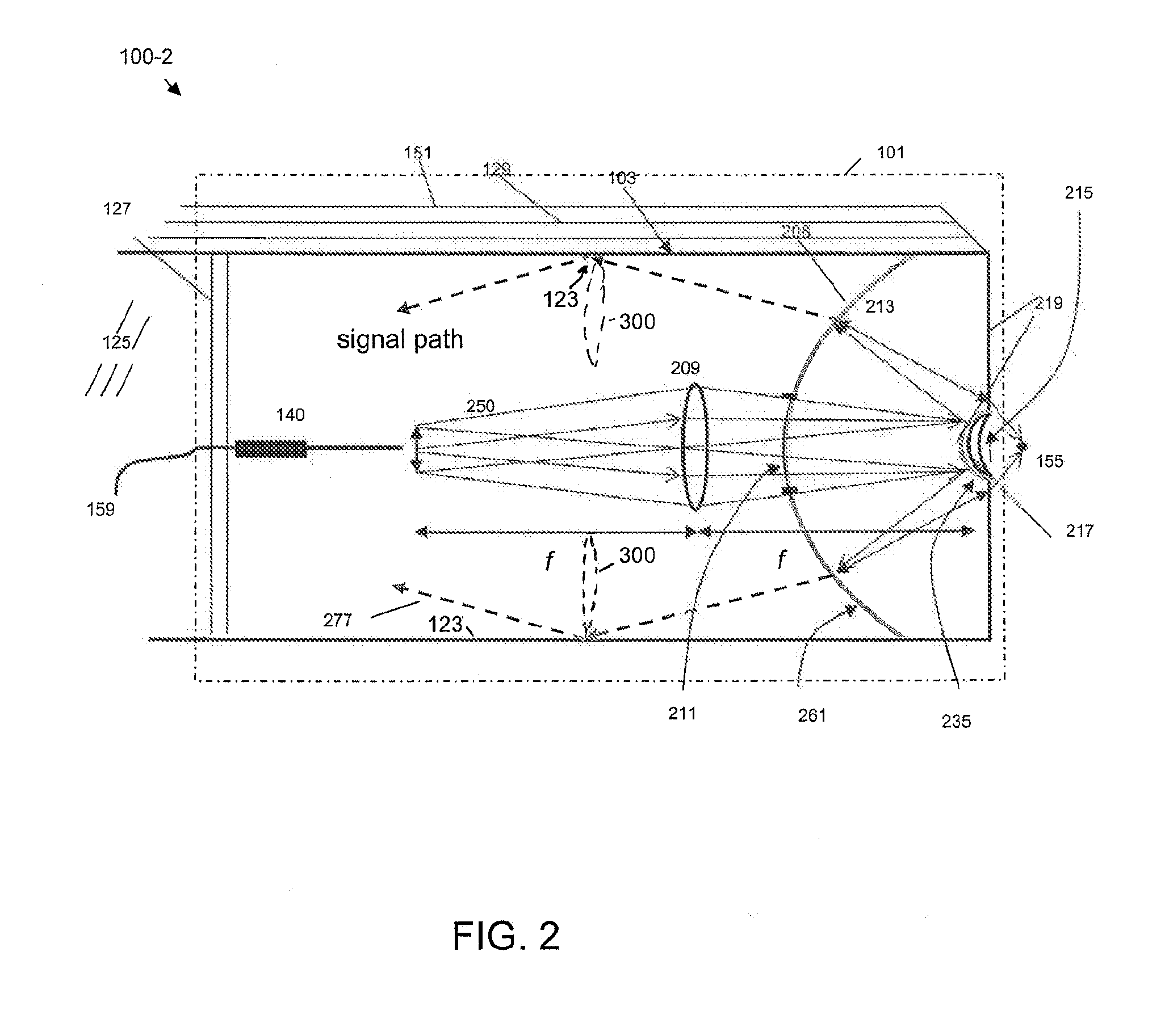
[0015]As used herein, the term “fluorescence emission” will be used to refer to multiphoton (particularly, two-photon but not excluding higher order) fluorescence emission as well as optical second harmonic generation (SHG) (but not excluding higher-order harmonic generation) from a target medium under conditions suitable to excite such fluorescence emission.

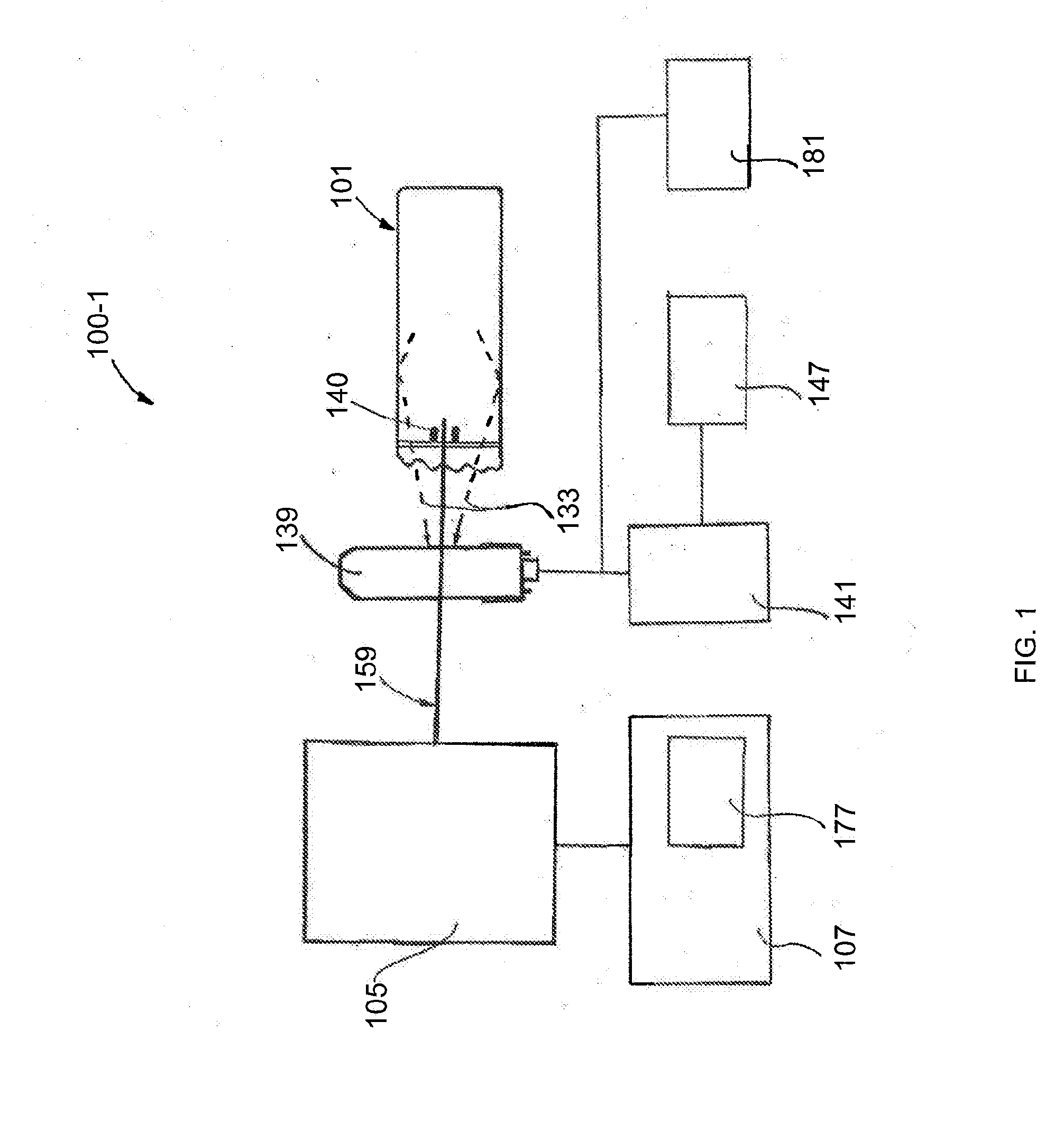
[0016]Illustrative embodiments of the invention include, but are not limited to, an optical system disposed in, or at, a distal end of a fluorescence emission endoscope, an optical system module for use in, or with, a fluorescence emission endoscope, an optical waveguide-based fluorescence emission endoscopy system, and a method for remotely-controlled, multi-magnification imaging of a target or fluorescence emission collection from a target with a fluorescence emission endoscope apparatus.

[0017]An embodiment of the invention is an optical system disposed in, or at, a distal end of a fluorescence emission endoscope apparatus. Th...


PUM


Abstract

Embodiments of the invention include an optical system and an optical system module, coupled to a distal end of a fluorescence emission endoscope apparatus, an optical waveguide-based fluorescence emission endoscopy system, and a method for remotely-controlled, multi-magnification imaging of a target or fluorescence emission collection from a target with a fluorescence emission endoscope apparatus. An exemplary system includes an objective lens disposed in a distal end of an endoscope apparatus. The lens is adapted to transmit both a visible target illumination and a fluorescence-emission-inducing target illumination as well as fluorescence-emission and visible light from the target. The system can thus simultaneously provide low magnification, large field of view imaging and high magnification, high-resolution multiphoton imaging with a single lens system.

Description

CROSS-REFERENCE TO RELATED APPLICATIONS[0001]This application claims priority to U.S. Provisional application Ser. No. 60 / 987,868 filed on Nov. 14, 2007, and to U.S. Provisional application Ser. No. 60 / 987,270 filed on Nov. 12, 2007, the subject matters of which are incorporated by reference herein in their entireties.STATEMENT REGARDING FEDERALLY SPONSORED RESEARCH OR DEVELOPMENT[0002]This invention was made with government support under Grant No. 1R01EB006736-01 and Grant No. 5-P41EB001976 sponsored by the National Institute of Biological Imaging and Bioengineering at the National Institutes of Health. The government has certain rights in the invention.BACKGROUND OF THE INVENTION[0003]1) Field of the Invention[0004]Embodiments of the invention are most generally related to the field of multiphoton fluorescence and / or non-linear harmonic emission endoscopy apparatus and methods. More particularly, embodiments of the invention are directed to multi-magnification, non-confocal, multi...

Claims


Application Information

Patent Timeline
Patent Type & Authority Applications(United States)
IPC IPC(8): A61B1/06A61B1/00
CPCA61B1/00165A61B1/043A61B5/0086A61B5/0071A61B5/0062A61B1/00172A61B1/00096A61B1/00188A61B1/0638A61B1/07G02B13/146G02B23/243G02B21/04G02B26/103
Inventor WEBB, WATT W.XU, CHRIS
Owner CORNELL UNIVERSITY
Why Eureka
Try Eureka