Flow cytometry detection method for mouse thymus or spleen T-lymphocyte subsets

A flow cytometry and lymphocyte technology, which is applied in the field of flow cytometry detection of mouse thymus or spleen T lymphocyte subsets, can solve the problems of large fluctuation and instability in the detection results, and achieve easy control and avoid errors. , the effect of simple operation steps

Inactive Publication Date: 2017-09-05
成都扬克斯生物科技有限公司
View PDF1 Cites 5 Cited by
  • Summary
  • Abstract
  • Description
  • Claims
  • Application Information

AI Technical Summary

Problems solved by technology

[0004] In view of this, the present invention provides a mouse thymus or spleen T lymphocyte Flow cytometry detection method of cell subpopulations, established a stable and standardized detection method of mouse thymus or spleen T lymphocyte subpopulations

Method used


Image

  • Flow cytometry detection method for mouse thymus or spleen T-lymphocyte subsets
  • Flow cytometry detection method for mouse thymus or spleen T-lymphocyte subsets
  • Flow cytometry detection method for mouse thymus or spleen T-lymphocyte subsets

Examples


Embodiment

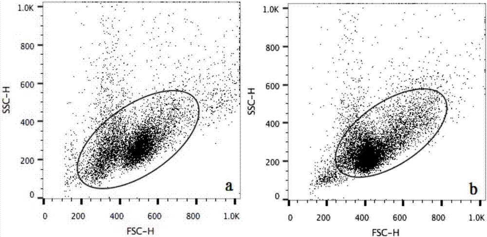
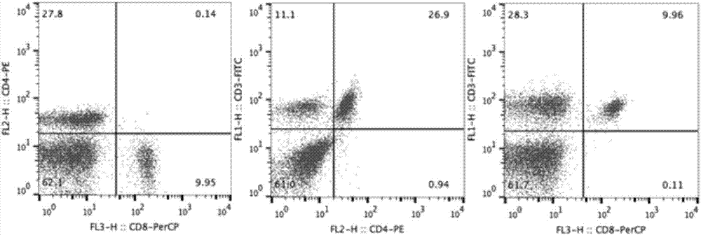
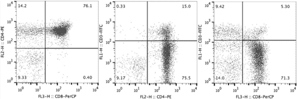
[0051] Step 1. Obtain the spleen or thymus tissue of the subject mouse and prepare a single cell suspension

[0052] According to a certain dose level, cyclophosphamide was injected intraperitoneally to the experimental mice for one week to establish an immunosuppressive model. The mice that were successfully modeled were sacrificed by cervical dislocation and killed immediately, the spleen or thymus tissue was taken out, the blood was blotted with filter paper, the surrounding connective tissue was trimmed with ophthalmic scissors, and stored in PBS at 4°C;

[0053] Take the preserved sample, put it in a watch glass, cut it repeatedly with ophthalmic scissors until the tissue becomes muddy, add an appropriate amount of PBS solution, and mix well; then filter it with a 300-mesh nylon mesh to obtain the filtrate; at a speed of 600-1000r / min Centrifuge the filtrate for 5 minutes, discard the supernatant; then add 1mL PBS solution, vortex to mix, centrifuge again, discard the sup...


PUM


Abstract

The invention discloses a flow cytometry detection method for mouse thymus or spleen T-lymphocyte subsets. The method comprises the following steps: acquiring thymus or spleen tissues of tested mice, and preparing single-cell suspension; respectively sucking the single-cell suspension of the thymus or spleen, respectively adding anti-mouse DC3, CD4 and CD8 monoclonal antibodies, swirling and uniformly mixing, keeping away from light at the temperature of 4 DEG C and staining; washing with PBS (Phosphate Buffer Solution) after staining, resuspending the cells, and detecting by using flow cytometry; and analyzing the detection result, thereby obtaining the percentage of each T-lymphocyte subset of the thymus or spleen of the mice. The stable and standardized method for detecting the mouse thymus or spleen T-lymphocyte subsets through flow cytometry is successfully established in the invention. By improving the problems such as target cell selection, compensative regulation and parameter selection in the sample pretreatment, detection and analysis process, errors caused by human factors in the test process are avoided, and the authenticity and objectivity of result judgment are improved.

Description

technical field [0001] The invention belongs to the technical field of cell detection, and in particular relates to a flow cytometry detection method of mouse thymus or spleen T lymphocyte subsets. Background technique [0002] Flow cytometry (Flow Cytometry, FCM) is a new technology for quantitative analysis and sorting of different types of cells according to the chemical composition of the surface or intracellular of a single cell. FCM mainly separates cells with different volumes by collecting forward scattered light (FSC), and collects side scattered light (SSC) to separate cells with different intracellular granularity. Furthermore, the detection of DNA, RNA, cytokines, cell surface antigens, etc. can be realized by identifying specific fluorescent markers on the cell surface and inside the cells. As a rapid cell analysis technology, this technology is not only used in basic medical research such as immunology and microbiology, but also widely used in clinical testing...

Claims


Application Information

Patent Timeline
Patent Type & Authority Applications(China)
IPC IPC(8): G01N15/14
CPCG01N15/1404G01N15/1434G01N2015/0065G01N2015/1006G01N2015/1415
Inventor 彭西于正强胡东李林蔚
Owner 成都扬克斯生物科技有限公司
Why Eureka
Try Eureka