Establishment method of solitary pulmonary nodule malignancy probability prediction model

A technique of probabilistic forecasting and establishment of methods, applied in the medical field, can solve problems such as no objective and quantifiable indicators, and achieve the effect of simple and easy-to-use models and high reference value

Active Publication Date: 2017-10-24
CHINA JAPAN FRIENDSHIP HOSPITAL
View PDF5 Cites 19 Cited by
  • Summary
  • Abstract
  • Description
  • Claims
  • Application Information

AI Technical Summary

Problems solved by technology

[0003] At present, the timing of further examination and even surgical intervention for SPN cases is mainly based on the physician’s clinical experience, which is greatly affected by subjective factors. Physicians with different professional directions often have different opinio...

Method used


Image

  • Establishment method of solitary pulmonary nodule malignancy probability prediction model
  • Establishment method of solitary pulmonary nodule malignancy probability prediction model

Examples


Embodiment 1

[0020] Embodiment 1 A method for establishing a predictive model for the malignant probability of solitary pulmonary nodules

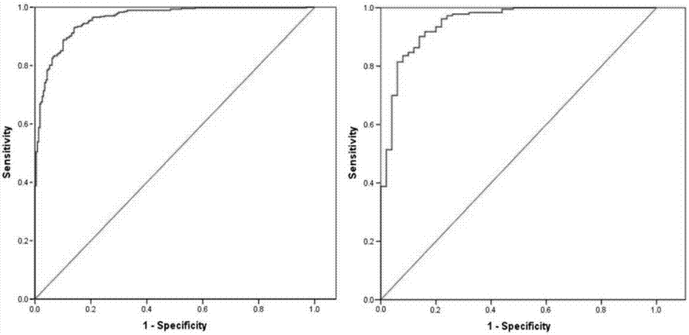
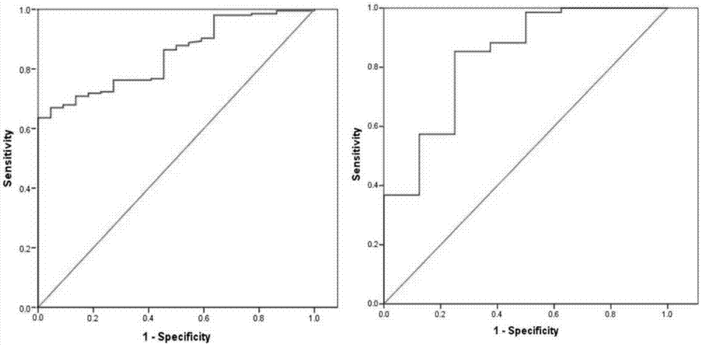
[0021] The applicant used 1234 cases of solitary pulmonary nodules, all of which had a definite pathological diagnosis. The general status of the patients was collected, including age, gender, smoking status (whether smoking, smoking amount, smoking cessation time, etc.), family cancer history, and past cancer history. Serum tumor marker levels were collected 1-7 days before surgery, including serum carcinoembryonic antigen (CEA) and serum bone collagen (Cyfra21-1) levels. Thoracic surgeons, respiratory physicians, and radiologists jointly reviewed and evaluated CT imaging features, including nodule location, CT maximum diameter (pulmonary window), proportion of ground-glass opacity (GGO), spiculation, Lobulation, calcification, cavity, well-defined borders, and pleural traction.

[0022] Considering that the difference in the proportion of nodules w...

Embodiment 2

[0029] Case 1: Male, 55 years old, physical examination found a nodule in the left lower lobe, the GGO component was less than 50%, the largest CT diameter was 1.8cm, no glitches, calcifications, pleural traction, no previous tumor history, and the serum tumor marker Cyfra21-1 value was 3.34 ng / mL, CEA value 2.82ng / mL.

[0030] Bring into the predictive model: P=e X / (1+e X), where X=-10.6+(0.064×age)+(1.358×largest diameter)+(1.698×burr)-(2.492×calcification)+(1.446×pleural traction)+(2.101×tumor history)+(0.639 ×Cyfra21-1)+(0.633×CEA). Calculated P=0.328, less than the cutoff value of 0.521.

[0031] After multiple consultations, the patient was finally operated on. The patient underwent pathological examination after the operation and was diagnosed as granulomatous inflammation. The possibility of old tuberculosis was considered, which was a benign nodule.

Embodiment 3

[0033] Case 2, male, 79 years old, physical examination found a right lower lobe nodule, GGO component greater than 50%, unclear border, serum tumor marker CEA value 5.33ng / mL.

[0034] Bring into the predictive model: P=e X / (1+e X ), where X=-1.180+(0.057*age)-(2.30*clear border)+(1.032*CEA). The calculated P=0.998 was greater than the cutoff value of 0.867.

[0035] On this basis, continued CT-guided puncture pathological diagnosis of lung adenocarcinoma, malignant nodules, received follow-up surgical treatment.


PUM


Abstract

The invention discloses an establishment method of a solitary pulmonary nodule malignancy probability prediction model. The establishment method particularly includes the steps: acquiring basic information of patients and serum tumor marker levels 1-7 days before operation; dividing patient cases into one group with GGO (ground glass opacity) lesion proportion higher than or equal to 50% and another group with GGO lesion proportion lower than 50% according to the GGO lesion proportion and CT (computed tomography) imaging reports of the patients; setting experiment groups and validation groups in each group of cases according to the proportion of 3:1, performing single-factor analysis on relative data of cases of the experiment groups to initially screen independent risk factors; substituting the independent risk factors into multifactor analysis to obtain independent risk factors for judging benign and malignant SPNs (solitary pulmonary nodules); acquiring the SPN malignancy probability prediction model by the aid of Logistic regression; substituting case data of the validation groups into the model, and verifying the case data of the validation groups. The model is simple and easy to use, used indexes can be acquired by the aid of routine examination and are easy to use, and effective intermediate reference information can be provided for further diagnosis and treatment of doctors according to the model.

Description

technical field [0001] The invention relates to the field of medical technology, in particular to a method for establishing a malignant probability prediction model for solitary pulmonary nodules. Background technique [0002] At present, it is generally accepted in the academic community that a round imaging nodule with a diameter of less than or equal to 3 cm and surrounded by lung tissue is defined as a solitary pulmonary nodule (SPN). According to literature statistics, about 1 in every 500 chest X-rays (0.2%) may show SPN, and 90% of SPNs are unintentionally discovered in physical examinations of healthy people. In the United States, more than 150,000 SPNs can be found in physical examinations every year. Common pathological findings of SPN include benign nonspecific granuloma, inflammatory granuloma, hamartoma, sclerosing hemangioma, etc., as well as malignant primary lung cancer and pulmonary metastases. There have been large-scale, prospective clinical studies in th...

Claims


Application Information

Patent Timeline
IPC IPC(8): G06F19/00
Inventor 肖飞梁朝阳
Owner CHINA JAPAN FRIENDSHIP HOSPITAL
Why Eureka
Try Eureka