Method for determining distal femur cuneiform plane in digital auxiliary mode

A femoral and distal technology used in the medical field to reduce surgical risks, shorten learning time, and reduce trauma

Inactive Publication Date: 2017-10-27
SICHUAN UNIV
View PDF0 Cites 19 Cited by
  • Summary
  • Abstract
  • Description
  • Claims
  • Application Information

AI Technical Summary

Problems solved by technology

Stulberg believes that this is caused by the inherent limitations of traditional mechanical positioning instruments, and the operator cannot obtain high accuracy by manually touching anatomical landmarks to locate the osteotomy

Method used


Image

  • Method for determining distal femur cuneiform plane in digital auxiliary mode
  • Method for determining distal femur cuneiform plane in digital auxiliary mode
  • Method for determining distal femur cuneiform plane in digital auxiliary mode

Examples


Embodiment Construction

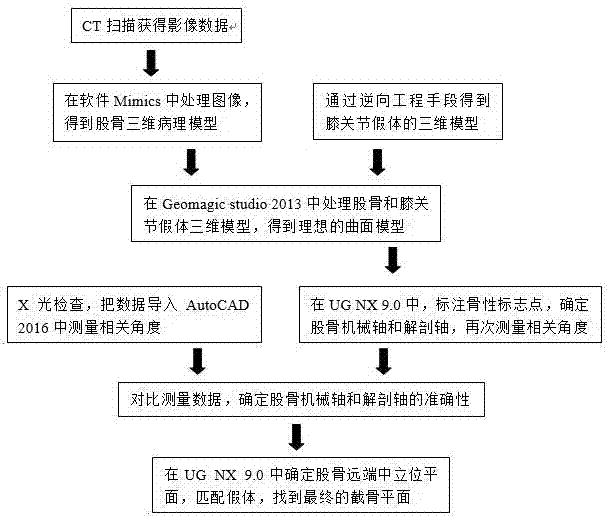
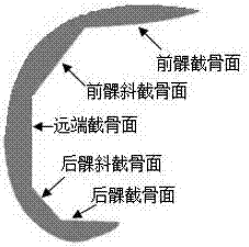
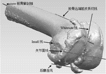
[0036] Below in conjunction with accompanying drawing, specifically illustrate the method that digitalization assists to determine the osteotomy plane of the distal end of femur:

[0037] 1. Obtain a structurally complete three-dimensional pathological model of the femur:

[0038] 1.1 Continuous tomographic CT scanning is performed on the patient. The scanning position is the patient’s supine position and lying down naturally, the knee joint is naturally straightened, and the patella points directly upward to prevent errors caused by rotation. The scanning site is about 160mm from the articular surface of the distal femur. About 50mm down to the articular surface of the distal femur;

[0039] 1.2 Import the obtained image files in DICOM 3.0 format into the interactive medical image processing software Mimics, select the bone window mode for image processing, and input in lossless compression mode, because the minimum gray value of bone tissue is generally greater than 1250, wh...


PUM


Abstract

The invention discloses a method for digitally assisting the determination of the osteotomy plane of the distal femur, which mainly includes the following steps: (1) performing continuous tomographic CT scanning on the patient to obtain image data; (2) obtaining the knee joint prosthesis by means of reverse engineering (3) Perform polygon processing and precise surface processing on the 3D data of the femoral and knee joint prosthesis; (4) Mark and correct the bony landmark points in the computer-aided design software; (5) Determine the osteotomy plane Perform osteotomy rehearsal. The advantages of the present invention are: (1) use digital means before operation to determine bony landmarks, axis, prosthesis model and osteotomy plane, etc., which can better plan the operation path and design the operation plan; (2) do not need to open the The medullary cavity determines the osteotomy surface, reduces trauma, reduces blood loss and the probability of fat embolism; (3) computer-aided positioning, improves the accuracy of positioning, makes the operation effect better, and shortens the patient's recovery time.

Description

technical field [0001] The invention relates to the field of medical technology, has distinct characteristics of medical-industrial integration, and specifically relates to a digitally assisted method for determining the osteotomy plane of the distal end of the femur before total knee replacement surgery. Background technique [0002] Osteoarthritis (OA), also known as osteoarthrosis (osteoarthrosis), is a common rheumatic disease. Osteoarthritis affects walking, up and down stairs and other lower limb functions of elderly patients, and some seriously cause joint dysfunction, joint deformity, and even disability. [0003] For patients with severe osteoarthritis, the most radical treatment is through artificial knee replacement. In traditional knee arthroplasty, the operator mainly determines the osteotomy plane through intramedullary positioning, which not only increases the risk of fat embolism and infection, but also increases the operation time and operation risk. [00...

Claims


Application Information

Patent Timeline
Patent Type & Authority Applications(China)
IPC IPC(8): A61B34/10
Inventor 王素杨洋洋徐亮李凯
Owner SICHUAN UNIV
Why Eureka
Try Eureka