Rapid super-resolution blood flow imaging method

An imaging method and super-resolution technology, which can be used in blood flow measurement devices, ultrasonic/sonic/infrasonic diagnosis, medical science, etc. The effect of calculating speed and improving detection efficiency

Active Publication Date: 2017-11-21
PEKING UNIV
View PDF6 Cites 18 Cited by
  • Summary
  • Abstract
  • Description
  • Claims
  • Application Information

AI Technical Summary

Problems solved by technology

In the "Nature" science journal, Erico et al. used a special ultra-high-speed sampling ultrasonic device to increase the frame rate of ultrasonic signal acquisition to 500 frames per second, thus shortening the time for forming a super-resolution image to 150s. Strict requirements on the performance of ultrasound equipment are difficult to apply in clinical
The core of ...

Method used


Image

  • Rapid super-resolution blood flow imaging method
  • Rapid super-resolution blood flow imaging method
  • Rapid super-resolution blood flow imaging method

Examples


Embodiment Construction
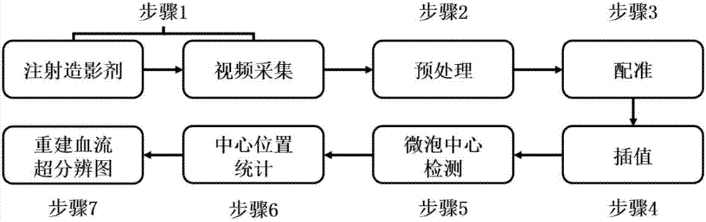
[0023] The idea of ​​the present invention is to collect ultrasonic images after contrast agent injection for preprocessing, interpolation, microbubble center detection, microbubble position statistics, and depiction of super-resolution images of blood flow. The following step diagrams and specific examples further illustrate the present invention, so that The invention is well understood, but the invention is not limited to this particular example.

[0024] figure 1 is the flow chart of the present invention for reconstructing super-resolution images of blood flow, such as figure 1 Shown:

[0025] In step 1, the model or individual to be imaged is first injected with a contrast agent, the imaging area is detected with an ultrasound probe, and data collection begins when a contrast-enhanced signal appears. The contrast medium can be given as a one-time injection or as a continuous injection. Different types of contrast agents and different imaging objects require different ...


PUM


Abstract

The invention discloses a rapid super-resolution blood flow imaging method based on an ultrasound microbubble contrast agent. By means of the method, the problem of low blood flow imaging resolution caused by an ultrasonic diffraction limit is mainly solved, and meanwhile super-resolution imaging is accelerated. The method includes the steps that 1, the contrast agent is injected, and ultrasonic video is collected; 2, the video is pretreated, and noise and background tissue are inhibited; 3, moving correction is conducted on the pretreated video; 4, image interpolation is conducted; 5, the center of a microbubble is computed frame by frame by means of a microbubble matching filter; 6, the central position of the microbubble in all frames is recorded; 7, the occurrence frequency of the microbubble in each pixel is counted, and a blood flow super-resolution image is formed by means of gray value. According to the rapid super-resolution blood flow imaging method based on the ultrasound microbubble contrast agent, the blood flow super-resolution image with the resolution exceeding that of traditional ultrasound contrast is formed in short time, more abundant blood flow details are provided for a doctor, and the method assists the doctor in clinical diagnosis.

Description

technical field [0001] The invention relates to the field of ultrasonic imaging systems and methods, in particular to a method for fast super-resolution ultrasonic blood flow imaging. Background technique [0002] The distribution of blood flow in the body and organs is a very common indicator used clinically. Now the main imaging methods are CT angiography (CTA), magnetic resonance angiography (MRA), digital subtraction angiography (DSA), and ultrasound Doppler (CDFI). CT-based angiography technology first injects CT contrast agent into blood vessels, and then performs CT imaging, which can distinguish blood vessels with a diameter of more than 400 μm. Magnetic resonance (MRI)-based angiography technology uses the fluidity of blood in the excited area to suppress the signal of immobile tissue, thereby highlighting the flowing blood. In the case of a field strength of 3.0T, blood vessels with a diameter of more than 300 μm can be distinguished. For fields above 3.0T Strong...

Claims


Application Information

Patent Timeline
IPC IPC(8): A61B8/06A61B8/00
CPCA61B8/06A61B8/481A61B8/5215
Inventor 张嘉宾龙云飞罗渝昆王月香张珏方竞文晶蒋文莉
Owner PEKING UNIV
Why Eureka
Try Eureka