
3D (Three dimensional) printed biological combination dental implant implanting device

A technology of 3D printing and dental implants, which is applied in dentistry, dental implants, dental restorations, etc., can solve the problems of high production cost, long diagnosis and treatment time, and unacceptable patients, and achieve low production cost, accurate operation, and simple structure Effect

Inactive Publication Date: 2017-12-01
CHANGSHA YUANDAHUA INFORMATION TECH CO LTD
View PDF2 Cites 2 Cited by
  • Summary
  • Abstract
  • Description
  • Claims
  • Application Information

AI Technical Summary

Problems solved by technology

Due to the complex internal conditions of the oral cavity, if only relying on the doctor's experience, the operation is likely to cause alveolar bone perforation, nerve tissue damage, or poor implant placement and angle.
[0004] However, to make a hole in the jawbone for implant insertion, there are nerves and blood vessels in the jawbone, and it is necessary to make a hole at a position determined by diagnosis. The hole is usually made using a so-called dental drill (commonly known as a turbine). For the appliance, the drill rod is installed on the front end of the dental drill for drilling. There are often problems: the drilling position cannot be found, the drilling is repeated, the hole is corrected, or when the first vibration occurs during drilling, The hole is larger than the implant, and the fit between the bone and the implant is loose, and sufficient initial fixation cannot be obtained, resulting in the failure of the implant itself. correct formation of the hole at the implant site in the
The reason for this is that in the CT image, the auxiliary guide plate must cover both the teeth and the soft tissue of the maxillary surface. The traditional image segmentation technology cannot separate the inner surface data of the auxiliary guide plate, so the basic shape of the guide plate cannot be reconstructed. , the diagnosis and treatment time is relatively long, the production cost is relatively high, and the process of pouring plaster is often difficult for patients to accept, it is not as good as a CT scan to obtain the inner surface model of the auxiliary guide plate

Method used


Image

  • 3D (Three dimensional) printed biological combination dental implant implanting device

Examples


Embodiment Construction

[0018] The specific embodiments of the present invention will be described in detail below with reference to the accompanying drawings. As a part of this specification, the principles of the present invention are explained through examples. Other aspects, features and advantages of the present invention will become clear through the detailed description. In the referenced drawings, the same or similar components in different drawings are represented by the same reference numerals.

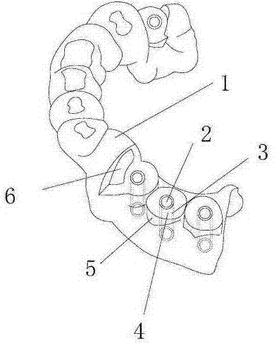
[0019] Such as figure 1 Shown here is a schematic structural diagram of the 3D printed biological composite dental implant device of the preferred embodiment of the present invention, the 3D printed biological composite dental implant device of the present invention, the dental implant device 1 completely fits the human teeth gingiva , The dental implant device 1 is provided with a drilling cap 5 at the missing tooth, a drilling platform 4 is provided on the drilling cap 5, a drilling guide hole 2 is ...


PUM


Abstract

The invention discloses a 3D (three dimensional) printed biological compound dental implant implanting device which is fully fitted with tooth gingiva of a human body. The dental implant implanting device is provided with a drilling sleeve cap on a tooth missed part, the drilling sleeve cap is provided with a drilling platform, a drilling guide hole is arranged in the center of the drilling platform, a rigid sleeve is arranged in the drilling guide hole, and air vents are arranged on the dental implant implanting device; the dental implant implanting device is printed by using a 3D printing technology. The 3D printed biological compound dental implant implanting device of the invention has the advantages of simple structure, reasonable design, convenient use and low production cost. According to the actual situation of different patients, different numbers and different diameters of the drilling guide holes are made, and a doctor can use the 3D printing technology according to different conditions of different people in the course of a surgery by utilizing the dental implant implanting device, so that the doctor can use a drill as advanced design each time in the surgery to enable the surgery to be more precise and scientific.

Description

Technical field [0001] The invention relates to a 3D printed biological composite dental implant device. Background technique [0002] 3D printing technology developed in 1987 to now, after more than 20 years, it has been rapidly developed and widely used worldwide. This method can quickly, accurately and personally manufacture computer models and CT scan data into implants with arbitrary shapes and structures. [0003] In dental implant surgery, the doctor uses a drill to drill the implant into the alveolar bone of the missing tooth. Due to the complex conditions inside the oral cavity, if you rely solely on the doctor’s experience, the operation is likely to cause perforation of the alveolar bone, nerve tissue damage, or poor implant placement and angle. [0004] However, a hole is made in the jawbone for implant insertion. There are nerves and blood vessels in the jawbone. It is necessary to make a hole at a position determined by diagnosis. The hole is usually called a dental d...

Claims


Application Information

Patent Timeline
Patent Type & Authority Applications(China)
IPC IPC(8): A61C8/00B33Y80/00
CPCA61C8/0012A61C8/0037A61C8/009A61C2201/00B33Y80/00
Inventor 郭志武
Owner CHANGSHA YUANDAHUA INFORMATION TECH CO LTD
Learn More