Kit and primer pair combinations for distinguishing active tuberculosis patients and non-tuberculous pneumonia patients

An active, tuberculosis technology, applied in the biological field, can solve problems such as low sensitivity, high laboratory biosafety requirements, and inability to meet clinical and tuberculosis prevention and control requirements

Active Publication Date: 2018-02-02
深圳市橙月生物科技有限公司
View PDF3 Cites 3 Cited by
  • Summary
  • Abstract
  • Description
  • Claims
  • Application Information

AI Technical Summary

Problems solved by technology

However, the current detection technology for diagnosing active tuberculosis is seriously inadequate and cannot meet the requirements of clinical and tuberculosis prevention and control
The gold standard for tuberculosis diagnosis is sputum Mycobacterium tuberculosis microbiological examination (sputum picture or sputum culture). Although this detection technique has high specificity, it has low sensitivity (less than 40%) and takes a long time (Tuberculosis culture takes 1- 2 months), the disadvantage of high laboratory biosafety requirements
The biggest disadvantage of this diagnostic method is that it cannot distinguish tuberculosis from other lung diseases caused by tuberculosis bacteria

Method used


Image

  • Kit and primer pair combinations for distinguishing active tuberculosis patients and non-tuberculous pneumonia patients
  • Kit and primer pair combinations for distinguishing active tuberculosis patients and non-tuberculous pneumonia patients
  • Kit and primer pair combinations for distinguishing active tuberculosis patients and non-tuberculous pneumonia patients

Examples


Embodiment 1

[0022] Example 1: Preparation of peripheral blood mononuclear cell (PBMC) suspension

[0023] Add 5ml of lymphocyte separation solution (Fresenius Kabi NOrge As: LYS3773) to the centrifuge tube; take 2ml of heparin anticoagulated venous blood from the above different types of people and mix the same amount of 1M phosphate buffer (PBS) thoroughly to obtain a mixed solution. Use a pipette to slowly superimpose the mixed solution on the lymphocyte separation liquid surface along the tube wall, keeping a clear interface, and centrifuge at 2000 rpm for 20 minutes; use a pipette to suck the middle cloud layer into another centrifuge tube and then add 5 Wash the cells with double volume of 1M PBS, 1500 rpm / dissociation for 10 minutes, discard the supernatant, and wash the cells again under the same conditions, then add 1ml of RPMI1640 (Thermo scientific: SH30807.01b) containing 10% calf serum volume percentage , Resuspend the cells to obtain PBMC suspension; in each case, take 20μl of P...

Embodiment 2

[0024] Example 2: RNA extraction

[0025] RNeasy Mini Kit (Cat. No. 74106) of Qiagene Company was used to extract RNA from the PBMC suspensions of the three groups of people obtained above. The specific operation is: take the above containing 1×10 6 The PBMC suspension of each cell is placed in a centrifuge tube with DNase and RNase removed at 3000 rpm / centrifugation for 10 minutes, and the supernatant is discarded; 350μl×Buffer RLT is added to the cell pellet, mixed well and lysed; 250μl absolute ethanol is added , Mix well, transfer the liquid to the RNeasy column, centrifuge at 8,000g for 30 seconds, discard the waste; add 350μl Buffer RW1 and centrifuge at 8,000g for 30 seconds, discard the waste; add 80μl DNase solution (10 μl DNase+70μlBuffer RDD) , Digest on the column for 15 minutes, centrifuge at 8,000g for 30 seconds, discard the waste; add 350μl Buffer RW1, centrifuge at 8,000g for 30 seconds, discard the waste; add 500μl Buffer RPE, centrifuge at 8,000g for 30 seconds...

Embodiment 3

[0026] Example 3: Reverse transcription

[0027] Using TAKARA's reverse transcription kit (DRR047), take 0.5μg of RNA obtained in step 2 for reverse transcription. Compared with traditional reverse transcription kits, this kit adds the steps of removing genomic DNA, which can guarantee to the greatest extent RNA purity and specificity of amplification.

[0028] The steps are as follows:

[0029] (1) Removal reaction of genomic DNA

[0030] Table 1 Removal reaction system of genomic DNA

[0031] Reagent

[0032] After preparing the reaction system according to Table 1, warm it at 42°C for 2 minutes, and the resulting RNA reaction solution from which genomic DNA has been removed is stored at 4°C.

[0033] (2) Reverse transcription reaction

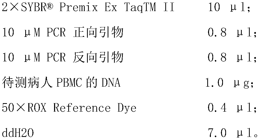
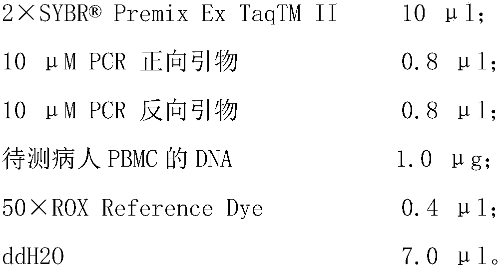
[0034] The preparation of the reaction system is carried out on ice, and the specific system is as follows:

[0035] Table 2 Reverse transcription reaction system table

[0036] Reagent

[0037] After preparing the reaction system according to Tabl...


PUM


Abstract

The invention discloses a kit and primer pair combinations for distinguishing active tuberculosis patients and non-tuberculous pneumonia patients. The primer pair combinations include SEQ ID NO:1-2, SEQ ID NO:3-4 and SEQ ID NO:5-6, wherein the SEQ ID NO:1-2 corresponds to a CD157 gene, the SEQ ID NO:3-4 corresponds to an IL-1b gene, and the SEQ ID NO:5-6 corresponds to an MS4A6A gene. By adoptionof the primers for RT-PCR expansion and substitution of related gene expression level into an established formula, the active tuberculosis patients and the non-tuberculous pneumonia patients can be distinguished, and accuracy and quickness in tuberculosis diagnosis are realized.

Description

Technical field [0001] The present invention relates to the field of biotechnology, in particular to a set of primer pair combinations and kits for distinguishing active tuberculosis patients from non-tuberculous pneumonia patients. Background technique [0002] Tuberculosis (Tuberculosis, TB) is a chronic infectious disease caused by Mycobacterium tuberculosis infection. Mycobacterium tuberculosis can not only cause tuberculosis (85%), but also cause tuberculosis in multiple organs outside the lungs. Although there are effective anti-tuberculosis drugs, tuberculosis is still the number one killer of current infectious diseases. About 2 million people die from tuberculosis every year. About one-third of the world’s population is infected with Mycobacterium tuberculosis. About 10% of people with latent tuberculosis infection (LTBI) will eventually progress to active tuberculosis. [0003] Due to the lack of effective tuberculosis vaccines, the prevention and control of tuberculosis...

Claims


Application Information

Patent Timeline
Patent Type & Authority Applications(China)
IPC IPC(8): C12Q1/6883C12N15/11
CPCC12Q1/6883C12Q2600/112C12Q2600/158
Inventor 杨帆刘小山
Owner 深圳市橙月生物科技有限公司
Why Eureka
Try Eureka