
A three-dimensional cell spheroid migration monitoring method based on microfluidic chip technology

A microfluidic chip, three-dimensional cell technology, applied in the field of cell biology research

Active Publication Date: 2021-10-15
DALIAN INST OF CHEM PHYSICS CHINESE ACAD OF SCI
View PDF6 Cites 0 Cited by
  • Summary
  • Abstract
  • Description
  • Claims
  • Application Information

AI Technical Summary

Problems solved by technology

At present, the use of microfluidic chips to monitor the migration kinetics of three-dimensional cell spheroids, and to observe the research and analysis of the process of three-dimensional cell spheroids migrating from a two-dimensional plane to a three-dimensional matrix is ​​still in a blank stage.

Method used


Image

  • A three-dimensional cell spheroid migration monitoring method based on microfluidic chip technology
  • A three-dimensional cell spheroid migration monitoring method based on microfluidic chip technology
  • A three-dimensional cell spheroid migration monitoring method based on microfluidic chip technology

Examples


Embodiment 1

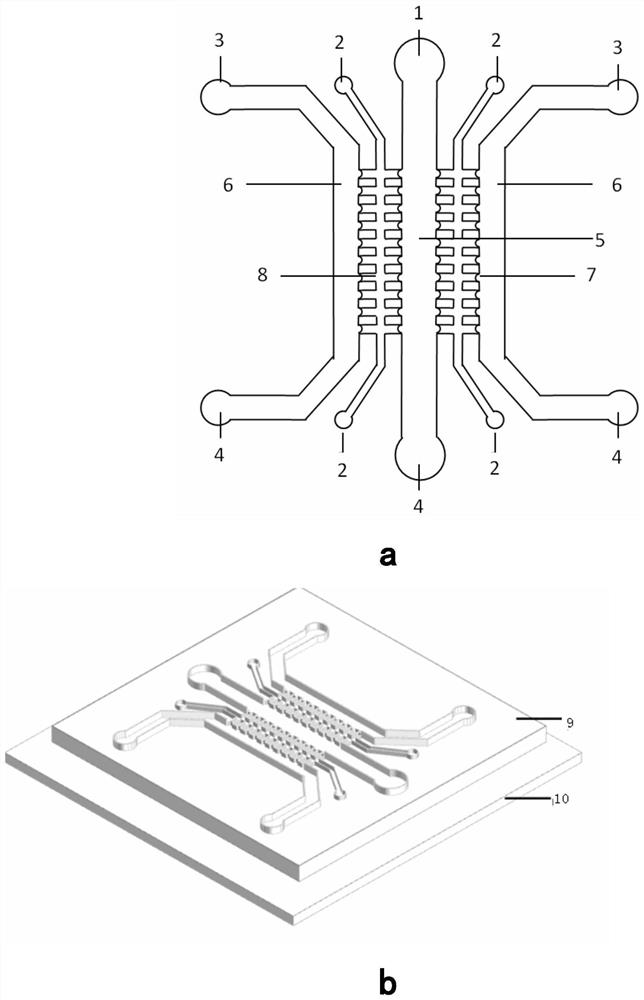
[0031] Using the microfluidic chip designed and produced by the laboratory, the configuration is shown in figure 1 . The microfluidic chip is composed of upper and lower layers of PDMS bonded and sealed, including cell inlet pool 1, collagen inlet pool 2, medium inlet pool 3, waste liquid pool 4, cell culture chamber 5, medium perfusion chamber 6, Cell ball capture tank 7, cell migration chamber 8;

[0032] The two ends of the cell migration chamber 8 are collagen inlet pools 2, the middle part is in the shape of "Feng", and the horizontal structure in the middle is 7 to 10 cell ball capture grooves 7 symmetrically arranged, and the cell migration chamber 8 passes through the cell ball capture grooves on one side 7 is connected to the medium perfusion chamber 6, and the cell migration chamber 8 is connected to the cell culture chamber 5 through the cell ball capture groove 7 on the other side;

[0033] The cell culture chamber 5 is connected to the cell inlet pool 1 on the u...

Embodiment 2

[0038] A three-dimensional cell spheroid migration monitoring method based on microfluidic chip technology, using the above microfluidic chip, according to the following steps:

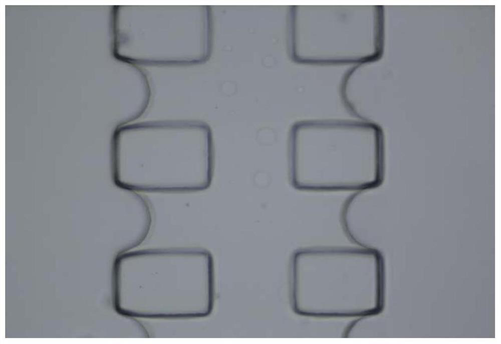
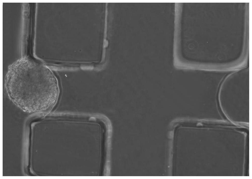
[0039] Prepare collagen with a concentration of 4mg / ml, pour it from the collagen inlet into the collagen channel of the chip, and incubate at 37°C for 30 minutes. After the collagen solidifies, you can see a clear semi-arc interface between the collagen and the two-dimensional plane, such as figure 2 shown. From the cell inlet pool at a lower density of 10 4 cm- 2 Add the three-dimensional cell sphere suspension, and stand the chip for 10 minutes to make the cells attach to the cell sphere capture area on the side of the collagen channel, such as image 3As shown, the cell channel and medium channel of the chip are filled with cell culture medium. Use the live cell workstation CO2 microscopic stage incubator for long-term observation of cells. Turn on the instrument, open the air and carbon dioxi...


PUM


Abstract

A three-dimensional cell sphere migration monitoring method based on microfluidic chip technology is provided, especially for the process of three-dimensional cell sphere migration into a three-dimensional matrix. The microfluidic chip is mainly composed of a cell inlet pool, a collagen inlet pool, a culture medium inlet pool, a waste liquid pool, a cell culture chamber, a medium perfusion chamber, a cell ball capture chamber, and a cell migration chamber. The method mainly includes the following steps: (1) chip collagen perfusion; (2) chip cell ball inoculation and culture; (3) chip cell migration real-time monitoring. The three-dimensional cell spheroid migration dynamics monitoring method based on the microfluidic chip has the characteristics of real-time tracking of cell movement, and at the same time can realize accurate positioning of the beginning of cell movement.

Description

technical field [0001] The invention relates to the technical field of applying microfluidic chip technology to real-time monitoring cell biology research, in particular to a microfluidic chip-based three-dimensional cell sphere migration monitoring method. Background technique [0002] Since the development of cell biology, the main culture and research have relied on orifice plates and commercialized transwell chambers, focusing on the observation of cell morphological changes, growth processes, migration and proliferation behaviors, etc. under the stimulation of single or multiple factors. In many cell behavior studies, cell migration, as the basis of important life processes such as organism development and morphogenesis, has attracted much attention. Cell migration generally begins with the cell's response to its microenvironmental stimuli, which activate a series of intracellular signal transduction pathways and gene transcription through cell surface receptors, and th...

Claims


Application Information

Patent Timeline
IPC IPC(8): C12Q1/02C12M3/00
CPCC12M23/16G01N33/5029
Inventor 秦建华陶婷婷李中玉张敏
Owner DALIAN INST OF CHEM PHYSICS CHINESE ACAD OF SCI
Learn More